3915
Accurate dynamic magnetic field monitoring and diffusion-weighted image reconstruction using uncorrelated local field measurements1Institute of Biomedical Engineering, National Taiwan University, Taipei, Taiwan, 2Department of Neuroscience and Biomedical Engineering, Aalto University, Espoo, Finland
Synopsis
We proposed a method to decouple field probes and to improve the accuracy of dynamic magnetic field estimation. A sensitivity matrix of probes was measured and to decouple NMR signals.
PURPOSE
Field probes can be used to estimate dynamic magnetic field map during MRI imaging. This information can be used to improve the image quality by, for example, mapping spiral trajectory1,2, estimating magnetic fields generated by cardiac and respiratory activity3, and tracking motion4. However, high quality probe measurements need to minimize the coupling between them in order to ensure localized and uncorrelated information. Signal couplings between field probes can result in incorrect dynamic magnetic field estimation, which eventually leads to erroneous image reconstruction. Note that incorrect dynamic magnetic field estimation attributed to coupled probes cannot be reduced by signal averaging.
Here we measured this signal coupling between field probes and proposed a strategy to mitigate this challenge. This decoupling method is based on a sensitivity matrix characterizing each probe to other probes. By using a strategy similar to sensitivity-encoded MRI5, we can separate the probe signal originating from its own droplet and minimize the coupled signals from other probes. To demonstrate our probe decoupling method, we used a 24-channel field probe array to monitor the dynamic magnetic field during diffusion-sensitive EPI measurements. Erroneously estimated magnetic field oscillation under constant gradient field was greatly reduced after applying the proposed probe decoupling method to generate dynamic magnetic map with up to the 4th-order spatial polynomials to improve the image reconstruction. Decoupled probes also led to reduced image artifact and improved image registration than from images using coupled probe signals.
METHODS
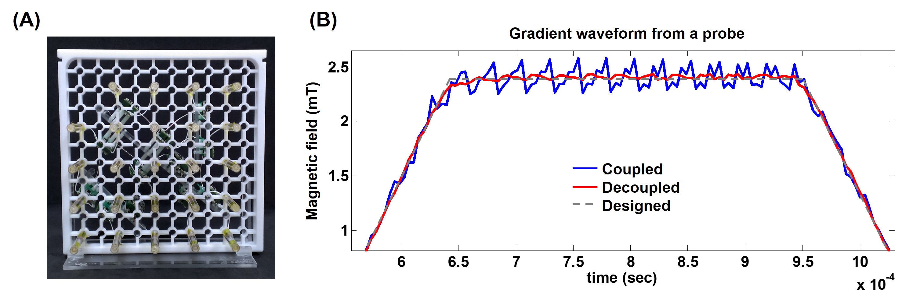
All scans were acquired on a 3T MRI scanner using a 20-channel head coil array (Skyra, Siemens Medical Solutions, Erlangen, Germany). Twenty-four field probes were arranged into a two-dimensional grid structure (Figure 1A). Diffusion weighted images (TR=6400ms, TE= 75ms, flip angle=90o, b=1000 s/mm2, three orthogonal diffusion sensitive gradient directions, resolution=3x3x4 mm3) were acquired using 20-channel head coil array and 24-channel field probes in separate sessions. All probes were distributed over the imaging plane and their locations were denoted by $$$\left ( x_{k}, y_{k} \right )$$$. A sensitivity matrix $$$\mathbf{S}$$$ with element $$$S_{ij}$$$ was used to denote the sensitivity of probe $$$i$$$ to the droplet in probe $$$j$$$. Note that $$$S_{ij}\neq S_{ji}$$$. The decoupled probe signals $$$p$$$ can be recovered by $$$p=\mathbf{S}^{-1}\tilde{p}$$$, where $$$\tilde{p}$$$ was a column vector denoting the collected coupled probe signals.
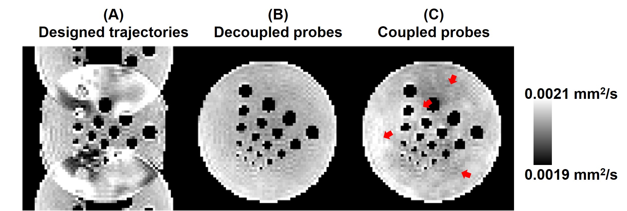
Images were reconstructed using (i) the designed trajectory without any correction, (ii) the actual magnetic field estimated by coupled probes, and (iii) by decoupled probes. Probe data were fitted up to the 4th-order spatial polynomials in magnetic field estimation.
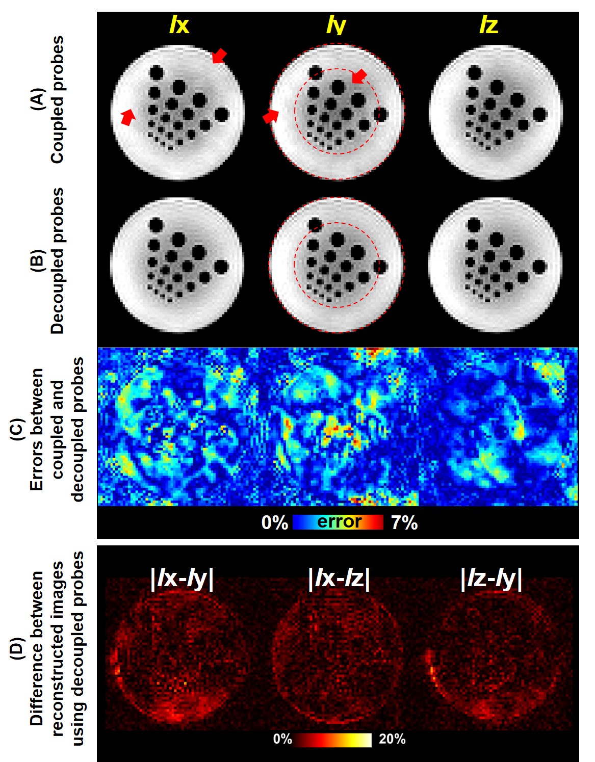
We used apparent diffusion coefficient (ADC) maps with diffusion sensitivity gradients in x, y, and z directions to test the performance of correcting artifacts caused by off-resonance using decoupled probes. Specifically, phantom images should have the same structure regardless of the applied diffusion sensitive gradients, which typically cause prominent and distinct off-resonance artifacts.
RESULTS
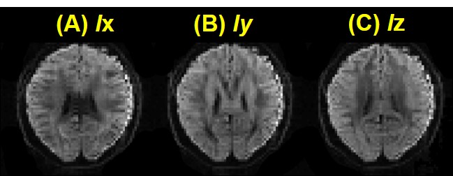
Figure 1B shows gradient waveforms of an EPI readout without diffusion sensitive gradient estimated by coupled and decoupled probes. Compared to the designed waveform, the erroneous oscillatory gradient waveform estimated by coupled probe was about six times larger than that estimated by decoupled probe. Ix, Iy, and Iz were images with diffusion-sensitive gradients applied to x, y, and z directions. Figures 2A and 2B are corrected images using coupled and decoupled probes. Different off-resonance artifacts were found among Ix, Iy, and Iz (Figure 2D), while these three images were very similar among one another using decoupled probles. Compared to coupled field probes, the image reconstruction error using decoupled field probes was reduced to 74%, 72% and 62% for |Ix – Iy|, |Iy – Iz| and |Iz – Ix| respectively. The reconstructed image without any correction showed serious ghosting artifact (Figure 3A). The ADC phantom image was homogenous using decoupled probes (Figure 3B), in contrast to the inhomogeneous ADC phantom image using coupled probes (Figure 3C). The in vivo reconstructed brain images with three different directional diffusion sensitive gradients, using magnetic field estimated from decoupled field probes, were shown in Figure 4.DISCUSSION
The coupling among probes resulted in signals mixture from different probes. The mixed probe signal had different resonance frequencies (due to different probe locations), which led to incorrect magnetic field estimation, artifacts and distortion. Probe coupling is a systematic error caused by the RF system and exists regardless of the chosen NMR-active signal source inside probes. However, this coupling can be easily removed by our proposed method and only needs to be measured once.Acknowledgements
This work was partially supported by Ministry of Science and Technology, Taiwan (103-2628-B-002-002-MY3, 105-2221-E-002-104), and the Academy of Finland (No. 298131).References
1. Barmet C, De Zanche N, Pruessmann KP. Spatiotemporal magnetic field monitoring for MR. Magn Reson Med 2008;60(1):187-197.
2. Sipila P, Greding S, Wachutka G, Wiesinger F. H-2 Transmit-Receive NMR Probes for Magnetic Field Monitoring in MRI. Magn Reson Med 2011;65(5):1498-1506.
3. Vannesjo SJ, Wilm BJ, Duerst Y, Gross S, Brunner DO, Dietrich BE, Schmid T,Barmet C, Pruessmann KP. Retrospective Correction of Physiological Field Fluctuations in High-Field Brain MRI Using Concurrent Field Monitoring. Magn Reson Med 2015;73(5):1833-1843.
4. Ooi MB, Krueger S, Thomas WJ, Swaminathan SV, Brown TR. Prospective Real-Time Correction for Arbitrary Head Motion Using Active Markers. Magn Reson Med 2009;62(4):943-954.
5. Pruessmann KP, Weiger M, Scheidegger MB, Boesiger P. SENSE: sensitivity encoding for fast MRI. Magnetic resonance in medicine 1999;42(5):952-962.
Figures