3905
Assessment of Geometric Distortion in EPI with a SPAMM Tagged Acquisition1Department of Imaging Physics, The University of Texas M.D. Anderson Cancer Center, Houston, TX, United States
Synopsis
Diffusion weighted EPI is susceptible to distortions due to multiple causes, and the amount of distortion is dependent on many interacting factors. Hence no good methodology currently exists for assessing and characterizing these distortions. In this work an EPI sequence is modified to include Spatial Modulation of Magnetization (SPAMM), a preparation technique that produces tagged grid lines in the imaged volume. This sequence is acquired to measure induced distortion in several phantoms in different coils with different sequence options, measuring distortion as it varies with parallel imaging acceleration, diffusion weighting direction, and susceptibility of various phantom materials.
Introduction
Distortion in diffusion weighted EPI is induced by a variety of causes, including magnetic field inhomogeneity, subject susceptibility, and eddy currents in the MR system. The amount of observed distortion is also dependent on the subject as well as imaging parameters and sequence options. Despite being a clinically important sequence, no methodology currently exists for meaningfully assessing all the factors that generate these distortions. Running conventional DW-EPI in existing phantoms designed for assessing geometric accuracy may be sufficient to provide a simple measure of distortion, but may not be adequate to characterize distortion in all clinical scenarios. We propose a simple, multi-slice technique that can be applied broadly to a wide variety of phantoms and sequence variations, forming the basis for a method to QA of spatial distortion exhibited by EPI sequences. By creating landmarks in the sequence itself, phantoms can be designed to simulate a wider range of scenarios, enabling the investigation of the independent causes of distortion in any given clinical protocol, including subject-induced distortions.Methods
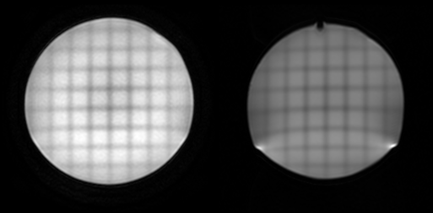
The proposed methodology utilizes a saturation technique known as Spatial Modulation of Magnetization (SPAMM) that tags the imaging subject with saturated grid lines1,2. Originally designed for tracking cardiac motion, these grids are applied here to assess spatial differences between diffusion weighting directions and/or b-values. For an absolute measurement, they can also be compared against a more geometrically robust sequence such as conventional fast spin echo. The saturation preparation sequence as implemented in this work consisted of binomially weighted (e.g. 1-3-3-1) pulses interleaved with gradient blips along the frequency encode direction, followed by the same sequence with gradient blips in the phase encode direction. Gradient area of the blips was adjustable and was typically set to produce line spacings of either 1.5 or 2.0 cm. Total time for the preparation sequence was about 9 msec.
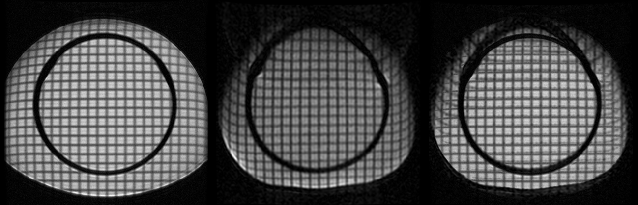
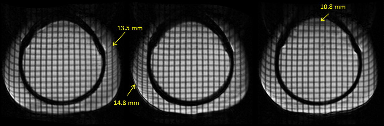
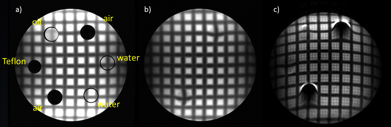
Three phantoms were assessed: a spherical head-sized phantom in an 8-channel head coil, a large shimming phantom in the quadrature body coil, and a modified PET ACR phantom that included compartments of water, air, oil, and Teflon, scanned in a quadrature head coil. Each phantom was acquired with three sequences using parameters from a clinically appropriate protocol: a conventional DW-EPI sequence (3 orthogonal diffusion weighting directions), the tagged DW-EPI with identical sequence parameters, and the tagged FSE sequence. Sequence parameters in the head phantoms were: FOV = 22, thickness = 4mm, matrix = 128x128, b-value = 1000, ASSET acceleration factor = 2 and 1 (separate acquisitions), TR=8000, TE=64.2. Sequence parameters for the large FOV shim phantom were: FOV = 46, thickness = 5, matrix = 128x128, b-value = 800, TR=4000, TE-57.3. The spherical head phantom was used to assess differences with ASSET acceleration, the large shim phantom was used to assess eddy current induced differences between diffusion directions, and the modified PET ACR phantom was used to assess differences in materials of varying susceptibility. Differences in grid locations were visualized with minimum intensity projection between images, and manually measured using intersecting locations on the grids.
Results
Grid lines were clearly visualized on tagged images and enabled quantification of distortions. On the spherical head phantom, parallel imaging acceleration was observed to improve distortion by up to 7 mm in areas close to susceptibility. On the large FOV shim phantom, maximum eddy current induced errors of 10.8 to 14.8 mm were observed in areas away from isocenter with diffusion weighting gradients applied in various directions. The modified PET ACR phantom demonstrated varying levels of distortion corresponding to the expected susceptibility of the materials in the compartments, including distortions of more than 2 cm near the air-water interface.
Discussion
SPAMM tagging provides a promising mechanism for assessing spatial distribution of distortion in EPI sequences. The preparation sequence is relatively short compared to the long TR EPI and FSE sequences, and places few if any limitations on the sequences. Since the tagged grid provides landmarks for spatial measurements, phantoms do not need to be designed with landmarks for spatial measurements, significantly expanding the types of distortion that can be assessed. These measurements can easily be extended to evaluate various sequence options and reconstruction or correction methods designed for DW-EPI sequences. The use of tagged grids may also be applied in patients for measurement or characterization of distortion in applications requiring high spatial accuracy such as treatment planning.Acknowledgements
No acknowledgement found.References
1. Zerhouni EA, Parish DM, Rogers WJ, Yang A, Shapiro EP: Human heart: tagging with MR imaging-a method for noninvasive assessment of myocardial motion. Radiology. 1988, 169: 59-63.
2. Axel L, Dougherty L: MR imaging of motion with spatial modulation of magnetization. Radiology. 1989, 171: 841-845.
Figures