3881
Penta-contrast imaging: a Novel Pulse Sequence for Simultaneous Acquisition of Proton Density, T1, T2, T2* and FLAIR images1Department of electrical and computer engineering, Seoul National University, Seoul, Korea, Republic of, 2Department of Radiology, Seoul Saint Mary's Hospital
Synopsis
A penta-contrast imaging sequence that generates proton density (PD), T1, T2, T2* and FLAIR images in a single scan is developed. Compared to conventional imaging sequences, the scan time is reduced by 50 %. Additionally, the new method generates T1 and T2 maps.
Introduction
In clinical MRI scans, multiple contrast images (e.g. T1-w, T2-w, FLAIR, …) are often acquired to obtain diagnostically valuable information. In brain imaging, PD, T1, T2, T2* and FLAIR are frequently acquired image contrasts. However, scan time for these five contrast images often takes more than 10 min. even when a thick 2D slice is obtained. Moreover, in certain applications, tissue parametric maps (e.g. PD, T1 and T2 maps) are acquired to measure pathology quantitatively or to observe longitudinal signal changes quantitatively. However, these parametric mapping methods requires even longer scan time because of repetitive acquisitions of multiple TRs and TEs. To accelerate the acquisition of MR data, several techniques that acquire multiple contrast images in a single scan have been reported.1-3 In this work, we introduce a novel pulse sequence that enables simultaneous acquisition of the five contrast images in less than 6 min. for the full brain. In addition, the proposed sequence provides T1 and T2 maps by utilizing the acquired images. The method may have potentials as a rapid evaluation tool for clinical scans.Methods
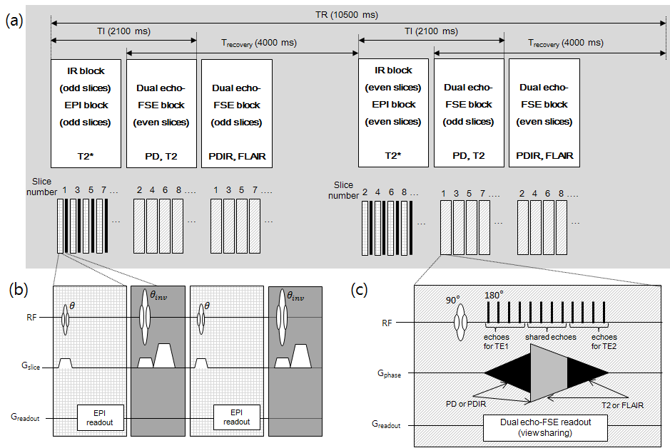
[Design of Penta-contrast sequence]
The schematic and timing diagram of the proposed pulse sequence is illustrated in figure 1A. In the penta-contrast sequence, the primary data acquisition unit was a dual-echo fast spin-echo (DE-FSE) to generate PD and T2 images simultaneously. Additionally, an inversion recovery RF was applied in every other DE-FSE acquisition to generate inversion recovery proton density (PDIR) and FLAIR images. Lastly, the gap between inversion pulses was filled with segmented EPI for T2* images (Fig. 1B). All of these were combined into a single sequence in a time-efficient way (Fig. 1) to reduce the scan time. For DE-FSE, the edges of k-space phase encoding lines were shared by PD and T2 images to further reduce the scan time.4 The slice acquisition was interleaved between odd and even slices. The timing of all sequence blocks was carefully designed for CSF saturation in FLAIR while acquiring 20 slices.
[Scan Parameters]
The penta-contrast sequence was designed to acquire the following imaging parameters: FOV = 230x230 mm2, resolution = 0.9x0.9 mm2, slice thickness = 5 mm, and number of slices = 20. To achieve these imaging parameters, the sequence timing was set to TR/Trecovery/TI = 10500/4000/2100 ms, TE1/TE2 = 7.9/86.9 ms, and matrix = 256x256. Echo train length (ETL) for DE-FSE was 12 with the first 4 echoes and the latter 4 echoes were assigned to center-part of phase encoding lines of PD and T2 contrast images, respectively. The rest 4 echoes from the center of ETL were assigned to edge-part of phase encoding lines and shared by the two contrasts (Fig. 1C). T1-w images were generated using $$(PDIR*PD)/({PDIR}^2+{PD}^2)$$ . For EPI, ETL was 8 and one navigator echo was acquired. For comparison, conventional sequences for individual contrast were acquired using the following parameters: PD images using FSE with TR/TE = 2800/9.6 ms, T1 images using SE with TR/TE = 510/9.6 ms, T2 images using FSE with TR/TE=4000/84 ms, T2* images using GRE with TR/TE = 600/20 ms, and FLAIR images using IR-FSE with TR/TE/TI = 9000/84/2500 ms. Total scan time of conventional sequences for the five contrasts was 13 min. All scans were done at 3T with a local IRB approval.
Results
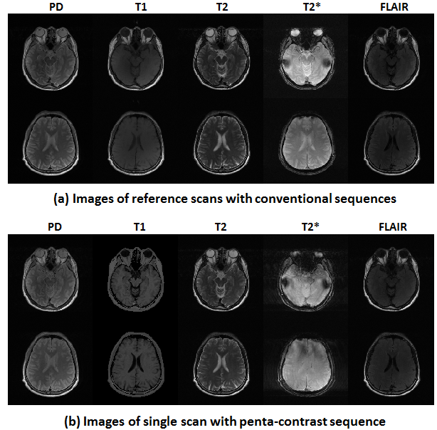
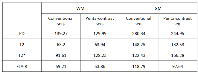
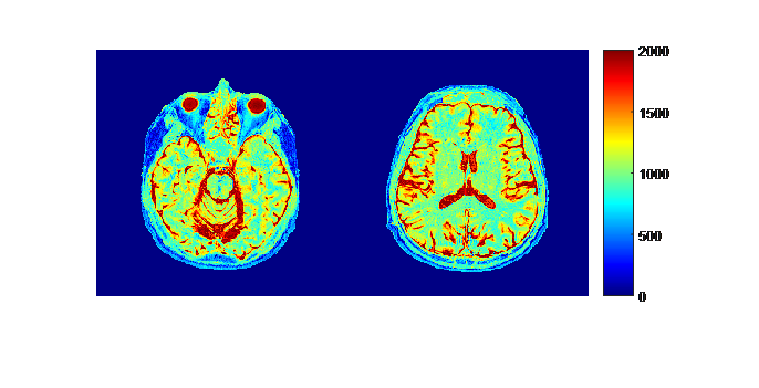
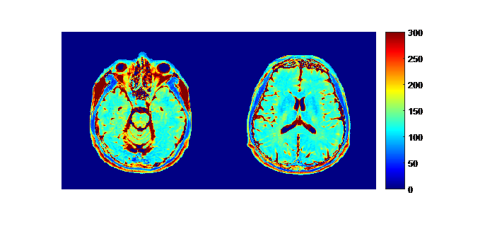
Figure 2 compares the images of the five different contrasts from the reference scans with the conventional sequences and the single scan with the proposed sequence. The penta-contrast images show similar contrasts to conventional images, while yielding a scan time reduction of 50%. The comparison of SNR in each contrast is summarized in Table 1. The SNR comparison shows similar SNR (T1 SNR is not included because the T1 image in penta-contrast was synthesized). The proposed sequence provides not only relaxation weighted images but also T1 and T2 maps as shown in Figures 3 and 4 which add important values.Discussion and conclusion
Here, we introduced a pulse sequence for simultaneous acquisition of the five contrast images. The results demonstrate the effectiveness of our approach for multi-contrast brain imaging. Additionally, T1 and T2 maps were generated at no cost providing additional benefits. Hence, the method may have potential applications as a fast evaluation tool for clinical scans. In our T2* weighted image, slight ghost artifacts due to phase variation between multi-shots were observed. This artifact may be alleviated by increasing ETL to reduce the number of shots. The estimated T2 maps show slightly over-estimated values which may originate from the stimulated echo in FSE. This overestimation may be corrected by using EPG.5Acknowledgements
This work was supported by the National Research Foundation of Korea (NRF) Grant funded by the Korean Government (No. NRF-2015M3C7A1031969) and by the Brain Korea 21 Plus Project in 2016.References
[1] Koichi Oshio and Ference A. Jolesz. Simultaneous Acquisition of Proton Density, T1 and T2 Images with Triple Contrast RARE Sequence. Journal of Computer Assisted Tomography 17 (2):333-338, March/April, 1993.
[2] Ralf Mekle, Andrew F. Laine, et al. Combined MR Data Acquisition of Multicontrast Images Using Variable Acquisition Parameters and K-Space Data Sharing. IEEE Transactions of Medical Imaging, Vol. 22, No. 7, July 2003.
[3] Kazuhiro Takeo, Akihiro Ishikawa, et al. FASCINATE: A Pulse Sequence for Simultaneous Acquisition of T2-Weighted and Fluid-Attenuated Images. Magnetic Resonance in Medicine 51:205-211, 2004.
[4] Blake A. Johnson, Evan K. Fram, et al. Evaluation of Shared-View Acquisition Using Repeated Echoes (SHARE): A Dual-Echo Fast Spin-Echo MR Technique. Am J Neuroradiol 15:667-673, Apr 1994.
[5] J Hennig, Multiecho Imaging Sequences with Low Refocusing Flip Angles. Journal of Magnetic Resonance 78, 397-407, 1988.
Figures