3876
Self-Calibrating Multi-Coil Phase Combination Using The Localized Singular Value Decomposition1Department of Imaging Physics, The University of Texas MD Anderson Cancer Center, Houston, TX, United States
Synopsis
MRI can encode clinically important information in the image-phase, thus has great potentials to extend its capability for clinical applications. However, if phase references are not available, the multi-coil phase combination using the weighted average suffers from dominant signal losses coming from phase cancellations due to uncompensated coil-dependent phase offsets.
In this work, we developed a self-calibrating multi-coil phase combining method which does not require any additional scans for phase references. When applied to a single-point Dixon imaging, we verified the proposed method could successfully estimate the global phase-map with the minimal signal loss only using a single-scan image.
Introduction
Magnetic resonance imaging can encode clinically important information in the image phase, thus has great potentials to extend its capability for clinical applications. In multi-channel MR phase imaging, time consuming multiple image acquisitions are required to remove both spatially varying coil-independent phase errors and coil-dependent phase offsets added to the clinically useful information in the image-phase. However, if phase references are not available, the multi-coil phase combination using the weighted average suffers from dominant signal losses coming from global and local phase cancellations due to uncompensated coil-dependent phase offsets. Due to this limitation, their clinical use has been restricted only when phase references are available using multiple image acquisitions1-2.
In this work, we developed a self-calibrating multi-coil phase combining method which does not require any additional scans for phase references. The proposed method uses the localized singular value decomposition (SVD) and rank-one approximation to estimate the initial global phase-map independently for each pixel, then the final multi-coil combined phase-map was composed after phase correction using a region-growing algorithm to estimate the unknown phase-signal polarity for each-pixel resulting from using localized SVD and rank-one approximation. The proposed method could successfully estimate the global phase-map with the minimal signal loss only using a single-scan image.
Methods
We developed a localized singular value decomposition (LSVD) method for multi-channel phase combination. Combining multi-coil phase-images using a single set of weighting coefficients calculated from the standard SVD is suboptimal especially when spatially varying coil-dependent phase offsets are uncompensated and inconsistent for multi-channel receivers. In our LSVD method, the initial global phase-map is estimated only using n x n surrounding neighboring pixels independently for each pixel. For each pixel, n x n neighboring pixels were rearranged in a [n2 x c] matrix where c is the total number of coils. Then, the [n2 x c] matrix of each pixel was decomposed using a standard SVD algorithm and the locally combined phase signals were estimated using the rank-one approximation (i.e. the largest singular value multiplied by the corresponding left singular vector)3. From the rearranged n x n image, only the central pixel was selected as the multi-coil combined signal for each pixel.
As the initial global phase map $$$e^{j\theta(\overrightarrow{r})}$$$ was estimated independently for each pixel using the localized SVD and rank-one approximation, the polarity of composed image-phase is unknown for each pixel and spatially inconsistent. Assuming that the image-phase is spatially smooth, the final phase-map was estimated by selecting a true solution between two candidates (i.e. $$$\pm e^{j\theta(\overrightarrow{r})}$$$) for each pixel using a region-growing algorithm4.
Results
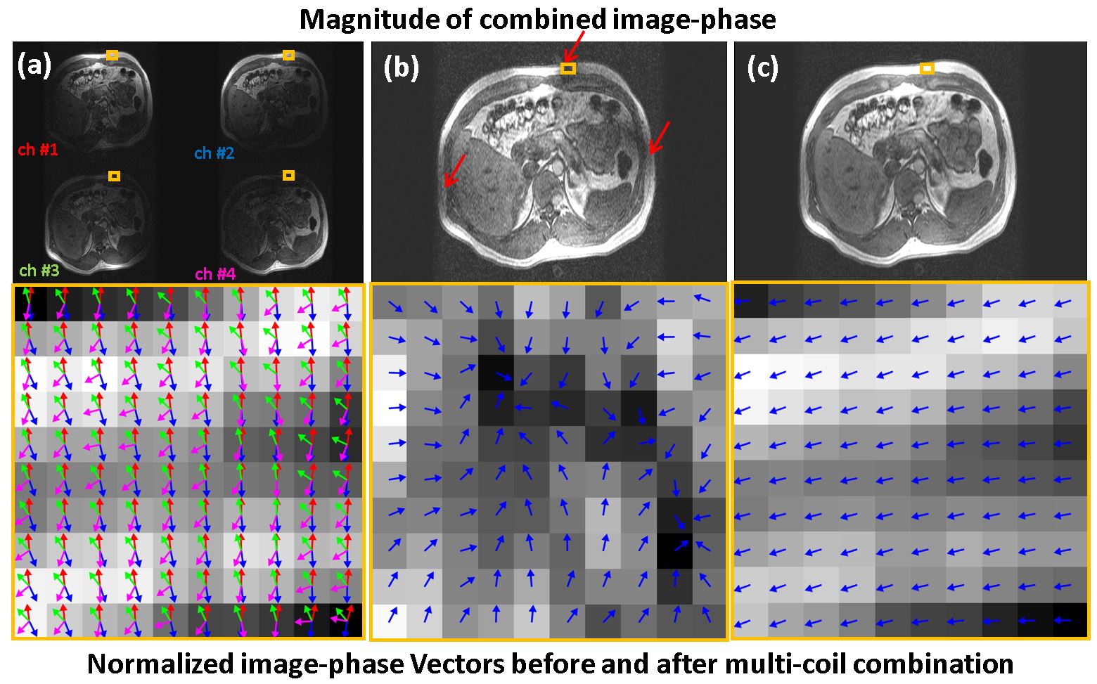
The self-calibrating multi-coil image-phase combining algorithm was tested using a single-point Dixon images5. The total of 32 in vivo abdomen images were acquired within a single breath-hold using the fast spoiled 3D gradient-echo pulse sequence on a 1.5T whole body clinical scanner (GE Healthcare, Waukesha) with 4-channel torso-phased array coils. Scan parameters are scan-time = 14 secs, TR/TE=4.2/1.668 ms, flip angle=15°, acquisition matrix=384x110, receiver bandwidth=±83.3 kHz, FOV= 36x24.75 cm, slice-thickness = 5mm, and phase-difference of water and fat signals=116°. The proposed algorithm was implemented in MATLAB (MathWorks, Natick). Fig. 1 (a) and (b) are normalized four-channel phase vectors and their weighted average in a selected region from the acquired single-point Dixon images (yellow boxes). Signal losses coming from the phase cancellation are clearly observed in the suboptimally combined image phase (red arrows) due to uncompensated coil-dependent phase offsets. On the other hand, the proposed method could successfully estimate the global phase-map with the minimal signal loss as shown in Fig. 1 (c).
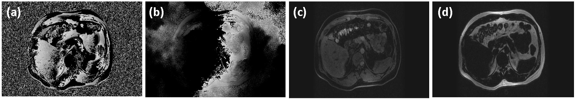
The initially estimated global phase map is shown in Fig. 2 (a). As the phase-map for each pixel was estimated independently for each pixel, the polarity of composed image-phase is unknown for each pixel and spatially inconsistent. Fig. 2 (b) is the final global phase-map estimated using simultaneously processed two phase-corrections using a single region growing algorithm4: (1) the phase correction to resolve the pixel-by-pixel polarity ambiguity resulting from using localized SVD and rank-one approximation, and (2) another phase correction required for Dixon imaging. After removing the estimated phase-map from the acquired single-point Dixon image, the uniformly separated water-only and fat-only images were reconstructed as shown in Fig. 2 (c) and (d).
Discussion
We verified the proposed method can successfully estimate the phase-map with the minimal signal loss only using a single-scan image when applied to an in vivo single-point Dixon imaging. Therefore, the propose technique has capability to help substantially expand the clinical use of MR phase imaging especially when the self-calibrating multi-coil phase combination is required.Acknowledgements
No acknowledgement found.References
1. Bernstein MA, Grgic M, Brosnan TJ, and Pelc NJ. Reconstructions of Phase Contrast, Phased Array Multicoil Data. Magn Reson in Med. 1994;32(3):330-334.
2. Lu K, Liu TT, and Bydder M. Optimal Phase Difference Reconstruction: Comparison of Two Methods. Magn Reson in Med. 2008;26(1):142-145.
3. Sandgren N, Stoica P, Frigo FJ, and Seléna Y. Spectral analysis of multichannel MRS data. Journal of Mag Reson 2005;175(1):79-91.
4. Son J, Jim XJ, and Ma J. 7. Three-dimensional T1-weighted MR imaging using a one-point Dixon technique with arbitrary echo time. In Proc of the 13th annual scientific meeting of Intl Soc Mag Reson Med, Miami, FL, USA, 2005:893.
5. Ma J. Breath-Hold Water and Fat Imaging Using a Dual-Echo Two-Point Dixon Technique With an Efficient and Robust Phase-Correction Algorithm. 2004;52:415-419.
Figures