3850
Ultrafast 2D MRI method based on multi-slice spatiotemporal encoding with simultaneous image refocusing1Department of Electronic Science, Xiamen University, Xiamen, People's Republic of China, 2Department of Communication Engineering, Xiamen University, Xiamen, People's Republic of China
Synopsis
We propose a new ultrafast multi-slice spatiotemporally encoded (SPEN) MRI technique, termed SeMSPEN, which outmatches conventional multi-slice EPI in its capability in shortening sampling time by producing multiple images in single echo train, and in lowering the specific absorption rate by segmenting the slice-selective dimension. The feasibility of this new method is verified theoretically and experimentally.
Purpose
To put forward a spatiotemporally encoded (SPEN) based multi-slice MRI sequence to reduce specific absorption rate (SAR) , shorten total scan time and improve image quality, while maintaining the advantage of SPEN in overcoming the effects of field inhomogeneity and chemical shift.Methods
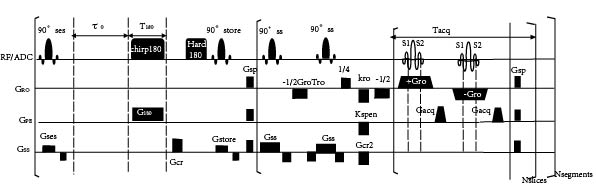
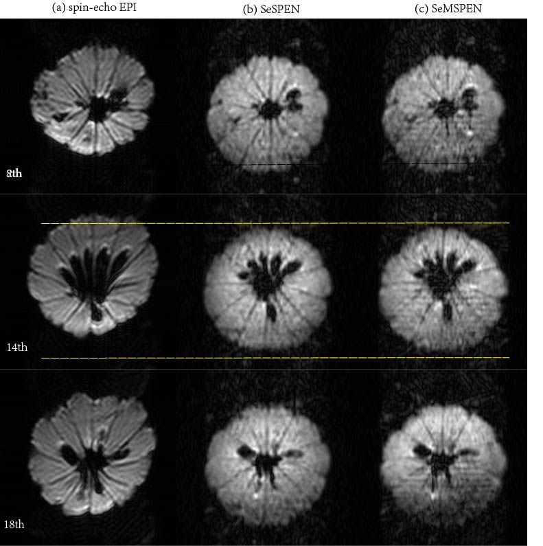
The new sequence, named SeMSPEN, is shown in Fig. 1. It initiates with a segment-selective 90° sinc pulse, which divides the whole sample volume into several segments. The ensuing 180° chirp pulse, targeting the full field of view (FOV) $$$L_y$$$ linearly, not only refocuses the magnetizations of excited slices, but also inverses the remaining longitudinal magnetizations. Then, a 180° hard pulse reverses all the spins not excited by the initial 90° segment-selective pulse. The quadratic phase evolves into: $${\varphi _1}\left( y \right) = {{\gamma {G_{180}}{T_{180}}} \over {{L_y}}}{y^2} + {{\gamma {G_{180}}{T_{180}}{L_y}} \over 4} - \gamma {G_{cr1}}{T_{cr1}}y$$where $$$\gamma$$$ stands for the gyromagnetic ratio, $$$G_{180}$$$ and $$$T_{180}$$$ the amplitude and duration of encoding gradient, $$$G_{cr1}$$$ and $$$T_{cr1}$$$ strength and duration of the first crusher gradient. A 90° sinc pulse is applied to restore all encoded information. Two signals from each slice are excited by two corresponding 90° slice-selective sinc pulses and acquired within a shared refocusing period. A specific linear combination of three dephasing gradients is employed to allow signals from different slices to refocus at different times on each readout period. When the areas of first, second and third dephasing gradients are set to -0.5, 0.25, -0.5 times of $$$G_{ro}T_{ro}$$$, respectively, the magnetization of the first slice (s1) experiencing 3 dephasing gradients will refocus at -0.75 $$$G_{ro}T_{ro}$$$, while the second slice (s2) experiencing 2 dephasing gradients will refocus at -0.25 $$$G_{ro}T_{ro}$$$. The final signal can be expressed as: $$s\left( t \right) \propto \int_{ - {{{L_y}} \over 2}}^{{{{L_y}} \over 2}} {\rho \left( y \right) \cdot {{ - \exp \left[ {i{\varphi _1}\left( y \right)} \right] - \exp \left[ { - i{\varphi _1}\left( y \right)} \right]} \over 2} \cdot \exp \left[ {i\left( {\gamma {G_{cr2}}{T_{cr2}}y + {k_{SPEN}}y + \gamma {G_{acq}}yt} \right)} \right]\exp \left( { - {\tau \over {{T_1}}}} \right)dy} $$where $$$\rho(y)$$$ represents the spatial profile of spin density, $$$k_{SPEN}$$$ is the initial purging gradient with an area of $$$|k_{SPEN}| = \gamma{G_{180}T_{180}}$$$, $$$\tau$$$ is the time between the 90° store pulse and the slice-selective pulse of the targeted slice ($$$90_{ss}$$$), and $$$T_1$$$ is the longitudinal relaxation time. To achieve full-decoding,1 the following conditions should be satisfied: $$$T_{acq} = 2{T_{180}}$$$, and $$$t_0 = T_{180}$$$. All experiments were performed on lemon samples on a 7T Varian MRI system equipped with a quadrature-coil probe. For comparison, spin-echo EPI and SeSPEN imaging2 were also performed. The parameters for acquisition were set as follows: FOV = 70 × 70mm2, matrix=128 × 64 (SeMSPEN) and 64 × 64 (EPI and SeSPEN), slice thickness = 1.5mm, frequency sweep bandwidth of chirp pulse = 4kHz (SeMSPEN) and 6kHz (SeSPEN), duration = 15ms (SeMSPEN) and 10ms (SeSPEN), spectral width = 500kHz, segment = 3, slices per segment = 8 (SeMSPEN sample two slices each time). Signals from SeSPEN and SeMSPEN were reconstructed and processed based on the de-convolution method.3
Results
Conclusion
The SeMSPEN method can remarkably reduce SAR and shorten total scan time while preserving the advantage of SPEN on resisting the effects of field inhomogeneity and chemical shift.Acknowledgements
This work was supported by the NNSF of China under Grants 11474236 and 81671674, and Science and Technology Project of Fujian Province of China under Grant 2016Y0078.References
1. Schmidt R, Frydman L. New spatiotemporal approaches for fully refocused, multislice ultrafast 2D MRI. Magn. Reson. Med. 2014; 71:711-722.
2. Zhang T, Chen L, Huang JP, Li J, Cai SH, Cai CB, Chen Z. Ultrafast multi-slice spatiotemporally encoded MRI with slice-selective dimension segmented. J. Magn. Reson. 2016; 269: 138-145.
3. Cai CB, Dong JY, Cai SH, Li J, Chen Y, Bao LJ, Chen Z. An efficient de-convolution reconstruction method for spatiotemporal-encoding single-scan 2D MRI. J. Magn. Reson. 2013;228:136-147.
Figures