3839
Simultaneous multi-slice triple-echo steady-state (SMS-TESS) T1, T2, PD, and B0 mapping in the human brain1Division of Radiological Physics, Department of Radiology, University of Basel Hospital, Basel, Switzerland, 2Department of Biomedical Engineering, University of Basel, Basel, Switzerland, 3Institute for Biomedical Engineering, University and ETH Zurich, Zurich, Switzerland
Synopsis
Triple-echo steady-state (TESS) has so far been investigated mainly as a particular robust and intrinsically B1-insensitive method for T2 relaxation time mapping. Here, its potential for fast simultaneous multi-parametric (T1, T2, PD, and B0) mapping of human brain tissues from a single scan is explored. TESS imaging is performed in 2D mode to mitigate motion sensitivity in the brain and accelerated by simultaneous multi-slice (SMS) imaging using CAIPIRINHA to excite two slices simultaneously providing similar SNR in half the acquisition time as compared to sequential single-slice imaging.
Introduction
Rapid simultaneous multi-parametric mapping of T1, T2, PD, and B0 based on triple-echo steady-state (TESS) imaging is investigated for human brain tissue characterization. TESS acquisitions are performed in 2D mode to reduce motion sensitivity and accelerated by a simultaneous multi-slice (SMS) excitation scheme.Methods
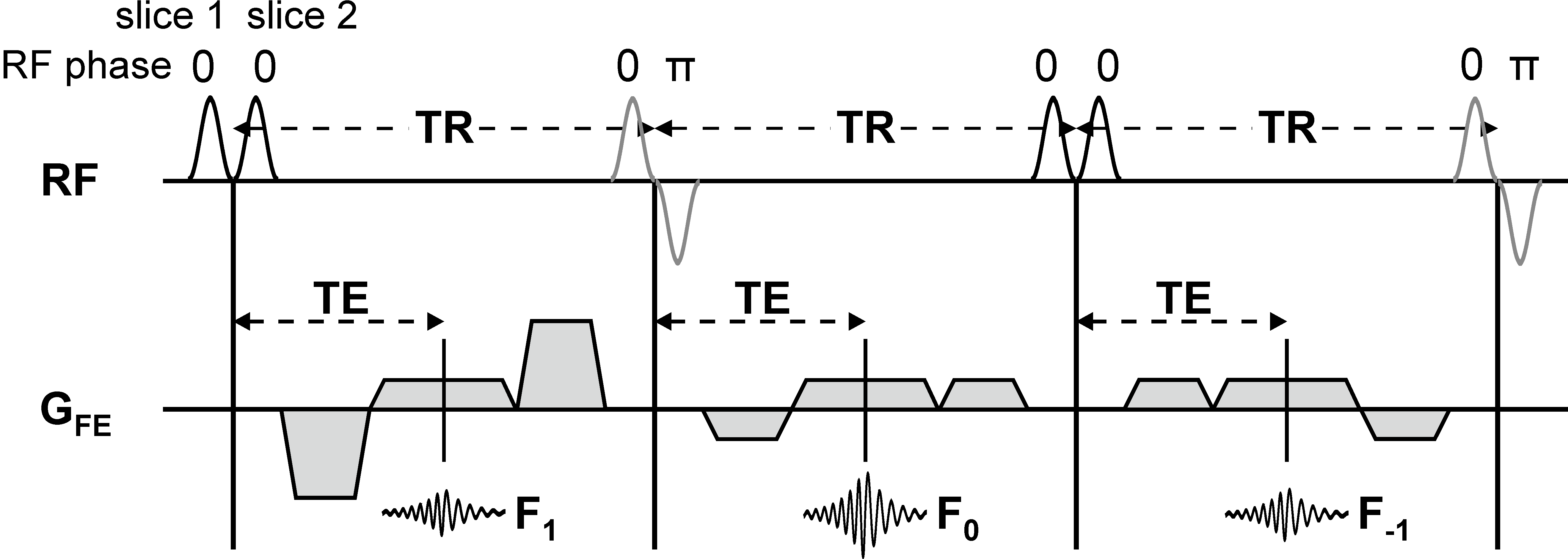
SMS-TESS imaging. A 2D TESS sequence1 is adapted for SMS imaging using a two-slice CAIPIRINHA excitation pattern2 (cf. Fig. 1). Imaging is performed in vitro in a manganese-doped aqueous probe (0.125 mM MnCl2 in H2O) and in vivo in the human brain at 3T with a standard 16-channel birdcage head coil. TESS protocol parameters are: TR = 9.84 ms, TE = 4.92 ms (in-phase), flip angle: 25°, receive bandwidth: 370 Hz/pixel, slice thickness: 4 mm, slice gap: 8 mm (edge-to-edge), in-plane resolution: (1.2 x 1.2) mm2 (matrix size: 208 x 156). Six averages are taken to provide sufficient SNR and mitigate motion issues (related to CSF pulsations), resulting in a total scan time of 28 s for two-slice CAIPIRINHA acquisitions (14 s per slice). TESS imaging is accompanied by a fast B1+ mapping scan of 8 s duration for T1 quantification3.
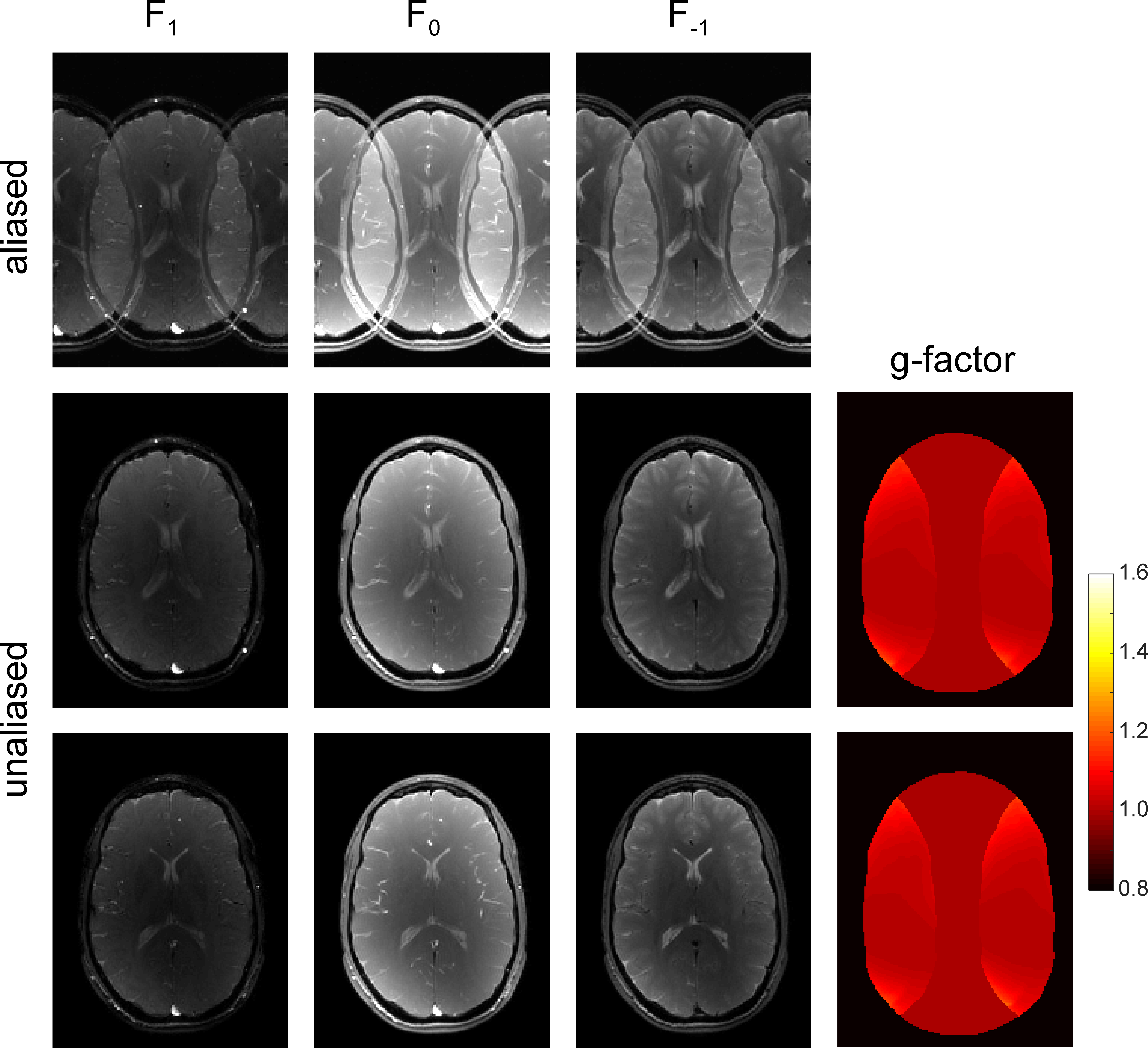
SENSE reconstruction. The aliased TESS images are reconstructed using SENSE4. The coil sensitivity maps required for the SENSE reconstruction as well as for the correction of the PD maps are estimated from a low-resolution spoiled gradient echo scan (TR = 4.9 ms, TE = 2.46 ms (in-phase), flip angle: 8°). The relative SNR compared to the unaccelerated case is calculated according to SNRCAIPIRINHA/SNRunaccelerated = 1/gCAIPIRINHA (gCAIPIRINHA: g-factor of the SENSE reconstruction)2.
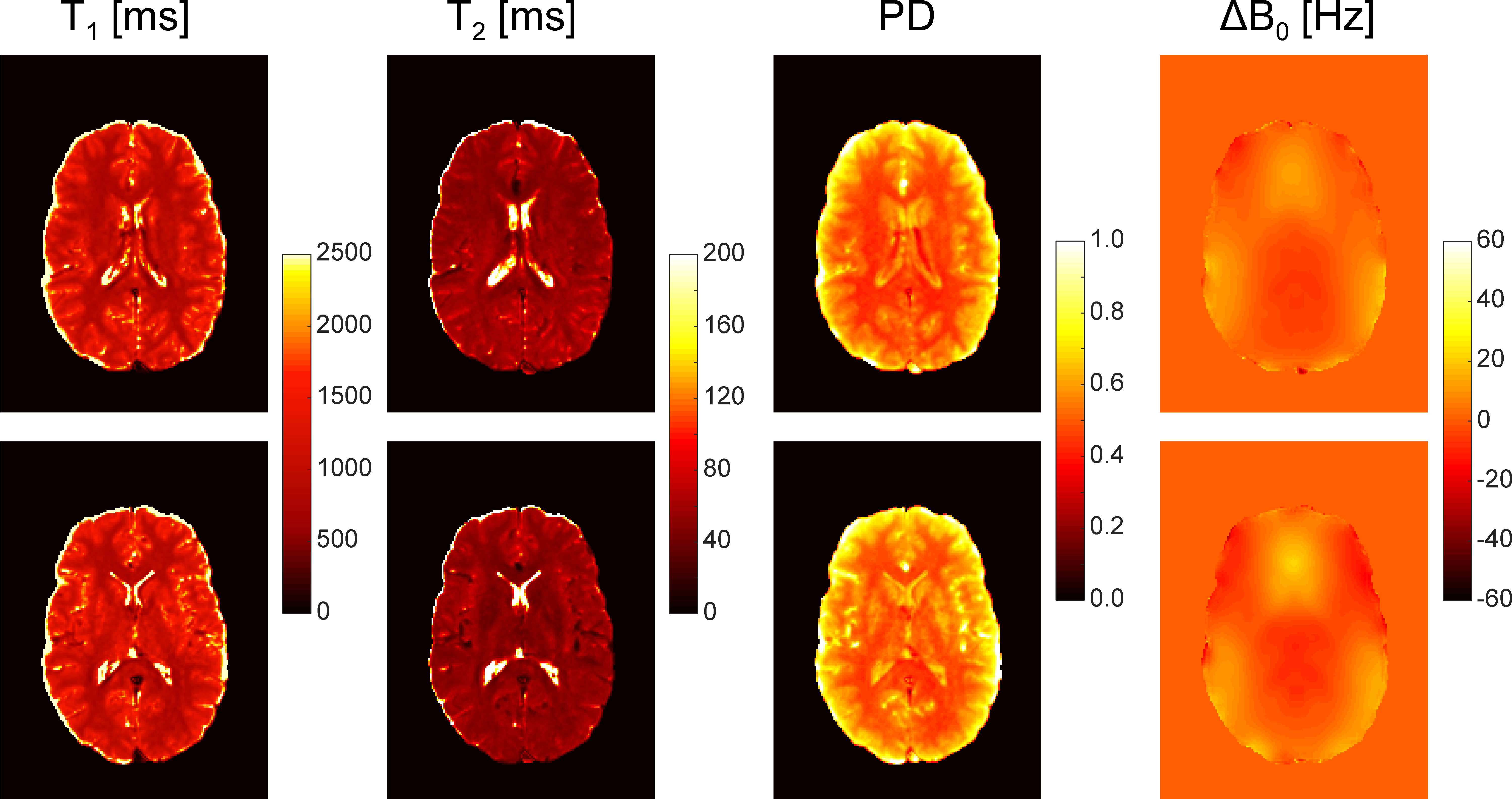
Multi-parametric mapping. The T1 and T2 relaxation times are determined according to the principle of 2D TESS relaxometry including B1+ and slice-profile correction1. PD maps are calculated based on the F0 signal amplitude by correcting the receiver coil sensitivity profile from the coil sensitivity calibration scan. Off-resonance frequency distribution (B0) is estimated by making use of the different phase evolution of the transverse F1 and F0 states. As the F1 state is refocused from the previous RF phase cycle with an effective echo time of TR+TE, the difference between the phases accumulated by the F1 and F0 configurations due to an off-resonance frequency Δω can be expressed as φF1 – φF0 = Δω∙TR giving a means for off-resonance frequency mapping with TESS.
Results
In vivo SMS-TESS imaging using two-slice CAIPIRINHA is demonstrated for human brain scans at 3T (cf. Fig. 2). The unfolded base TESS contrasts (F1, F0, and F-1) are of high quality without any visible reconstruction-related degradation (cf. Fig. 2, rows 2 and 3). A mean g-factor of 1.01 is obtained for both slices resulting in a mean relative SNR of 0.99 with respect to the unaccelerated case (see the corresponding g-factor maps in Fig. 2, last column). The T1, T2, PD, and B0 maps calculated from the SENSE-reconstructed TESS base images are shown in Figure 3 and prove the feasibility of multi-parametric TESS imaging in the human brain.
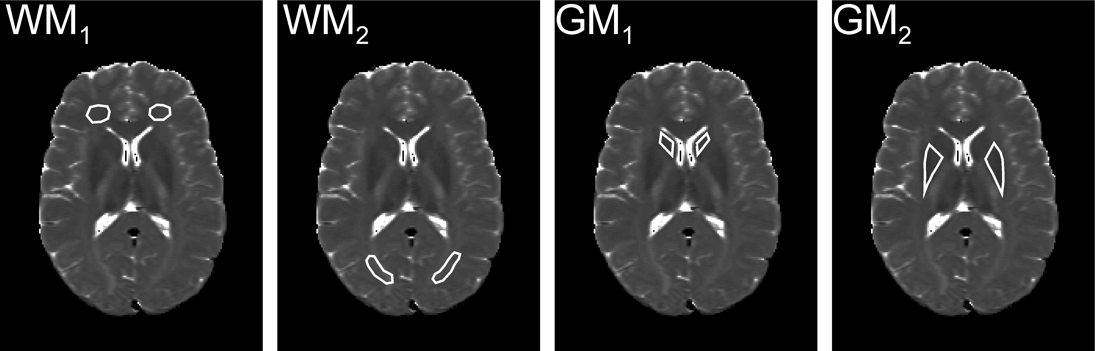
SMS-TESS T1 and T2 relaxometry validated against gold standard inversion-recovery (T1) and single-echo spin-echo (T2) measurements yields excellent agreement in vitro: 896 ± 18 ms (880 ± 2 ms) and 68 ± 1 ms (70 ± 1 ms) for T1 and T2, respectively (reference values given in brackets). In vivo, T1 and T2 accuracy is assessed for the regions-of-interest (ROIs) as indicated in Figure 4. Good agreement with the reference method is found for TESS-T1 in the white matter structures while in gray matter, a slight T1 overestimation is observed (cf. Table 1); possibly due to residual B1 inaccuracy. TESS-T2 yields overall good agreement with the spin-echo data (cf. Table 1).
Discussion
Using dedicated SMS imaging, 2D TESS acquisitions in the human brain can be accelerated by a factor of two while preserving the SNR (relative SNR of 0.99 compared to the unaccelerated case). While Hadamard excitation requiring multiple scans combined with water saturation pulses as recently proposed for 2D multi-slice TESS imaging in the brain5 is restricted to the accurate quantification of the T2 relaxation time, here, SMS-TESS combined with CAIPIRINHA reveals potential for rapid and accurate simultaneous mapping of T1, T2, PD, and B0 in the human brain. Future investigation may include to further accelerate TESS by using SMS-acceleration factors higher than two.Conclusion
SMS-TESS imaging with a CAIPIRINHA dual-band excitation pattern allows the rapid estimation of multiple tissue parameters (T1, T2, PD, and B0) based on the magnitude and phase information of three SSFP contrasts acquired in a single scan.Acknowledgements
This work was supported by a grant from the Swiss National Science Foundation (SNF 325230-153332).References
1. Heule R, Bar P, Mirkes C, Scheffler K, Trattnig S, Bieri O. Triple-echo steady-state T2 relaxometry of the human brain at high to ultra-high fields. NMR Biomed 2014;27(9):1037-1045.
2. Breuer FA, Blaimer M, Heidemann RM, Mueller MF, Griswold MA, Jakob PM. Controlled aliasing in parallel imaging results in higher acceleration (CAIPIRINHA) for multi-slice imaging. Magn Reson Med 2005;53(3):684-691.
3. Chung S, Kim D, Breton E, Axel L. Rapid B1+ mapping using a preconditioning RF pulse with TurboFLASH readout. Magn Reson Med 2010;64(2):439-446.
4. Pruessmann KP, Weiger M, Scheidegger MB, Boesiger P. SENSE: sensitivity encoding for fast MRI. Magn Reson Med 1999;42(5):952-962.
5. Pusterla O, Santini F, Heule R, Bieri O. T2-Snapshots imaging with simultaneous multislice TESS acquisition. Proceedings ISMRM 2015. p 441.
Figures