3830
SNR and volume characterization of RF coils: A simple procedure and an automatic post-processing tool for a straightforward comparison1Univ.Lyon, CREATIS, CNRS UMR 5220, Inserm U1206, INSA-Lyon, UJM- Saint Etienne Université Claude Bernard Lyon 1, Villeurbanne, France, lyon, France, Metropolitan, 2CERMEP – imagerie du vivant, Lyon, France, lyon, France, Metropolitan
Synopsis
To choose the most suitable RF coil available for an MRI study, we propose a procedure which uses a calibrated phantom, a 3D gradient-echo sequence, and an automatic post-processing tool available on the web. This tool generates a report which contains the measurement of a SNR with uniform volumes located in the depth. The post-processing could be done on MR images acquired on most main MRI vendors (Siemens, GE, Philips and Bruker) with prior verification of applied scaling or filtering. RF coil characterization results performed on at 4.7T and 7T were compared. The tool can be used for quality control.
Purpose
To propose a simple procedure and an available dedicated post-processing tool to characterize receiver RF coils in term of SNR and sensitivity volumes to assess and compare characteristics best suitable choice and quality control.Methods
Looking at the large number of RF coils available on MRI system, it could be difficult to make a choice for some unusual studies, whereas selecting the most suitable RF coil is critical to achieve the best MRI scan quality or to reduce acquisition time. The choice becomes even harder when several MRI systems from different vendors, operating at different static magnetic fields are available. It is the purpose of this procedure which uses a loaded phantom that mimics the biological conditions, a widely available 3D gradient-echo sequence with a short acquisition time (4min), and a post-processing available in free access at the web portal (no local installation necessary) [1]: ˝https://vip.creatis.insa-lyon.fr˝. The automatic post-processing generates a report file including:
(1) The SNR measurement corrected by some sequence parameters and the relaxation time based on the absolute SNR of McRobbie et al. [2] and adapted to a 3D gradient-echo sequence. To ensure comparable results, the program checks if image scaling [3] or filtering is applied to the image by using two statistical tests based on the noncentral-chi distribution of the noise in the background of the image [4].
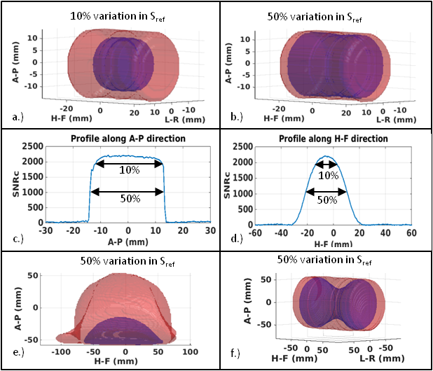
(2) 3D rendering of uniform volumes at 10% and 50% of tolerance as well as the capture zone of the RF coil defined at 90% of signal variation (see figure 1). These shapes with the addition of a table summarizing the characteristic lengths and intensity profiles in the three directions of the space allow us to evaluate the efficiency zone of the coil.
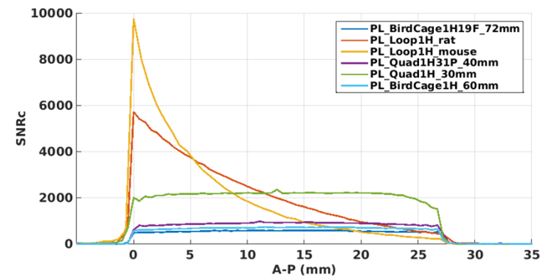
(3) An array of relevant measurements to compare the set of RF coils followed by a figure of the superimposed profiles inquired in SNR (see figure 2). The latter figure demonstrates the efficiency of a coil compare to the other by their intersection probed in the depth.
Results
The results shown in figure 2 were obtained on a 4.7T preclinical Bruker MRI system and with a phantom that mimic the body of mouse. For example, to scan the liver of a mouse, we demonstrated that the surface coil named 'PL_loop1H_rat' is the most efficient for a spectroscopic study, whereas the volume coils named 'PL_quad1H_30mm' give a better uniform response all over the organ with10% uniformity.
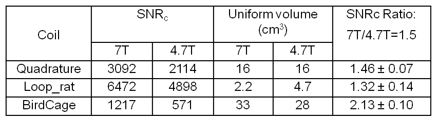
Acquisitions with the same phantom have been done on a 7T preclinical Bruker MRI system and have been compared to the latter results in a multicentre framework (see table 1). The results reveal similitude in the case of coils of identical constructor and different results between coils from different constructors. We noted that Bruker and GE MRI systems plot the original data of the acquisition when no filter is selected, whereas Siemens and Philips MRI systems rescale it automatically to make the images more attractive. This as a significant impact on measured SNR results. Reconstructions are required to retrieve the original data. When the acquisitions are usable, the uncertainty of the SNR was estimated at 5% in a reproducibility study, and 2% in a repeatability study.
Beyond to characterize the RF coils, this tool can be used as a quality control of the entire RF chain. The values of SNR and uniform volumes are saved after each post-processing. A new figure appears with the previous results, allowing a quality control throughout the time on the RF coil plus MRI system.
Conclusion
We propose a simple procedure and an automatic post-processing tool to characterize the RF coils in term of SNR and uniform volumes. Neither MRI sequence development nor software installation is required. The tool checks if scaling or filtering is applied on the image. The comparison of the results helps the user to evaluate the most suitable RF coil and MRI system for a specific acquisition. This tool can be used as a quality control thanks to the low uncertainty of the SNR measurement.Acknowledgements
This work was supported by France Life Imaging (FLI) and was performer within the framework of LABEX PRIMES (ANR-11-LABX-0063) of Université de Lyon, within the program "Investissements d'Avenir" (ANR-11-IDEX-0007) operated by the French National Research Agency (ANR).References
1. T. Glatard, C. Lartizien, B. Gibaud, R. Ferreira da Silva, G. Forestier, F. Cervenansky, M. Alessandrini, H. Benoit-Cattin, O. Bernard, S. Camarasu-Pop, N. Cerezo, P. Clarysse, A. Gaignard, P. Hugonnard, H. Liebgott, S. Marache, A. Marion, J. Montagnat, J. Tabary, and D. Friboulet. A Virtual Imaging Platform for Multi-Modality Medical Image Simulation. IEEE TRANSACTIONS ON MEDICAL IMAGING, 32, 110, 2013.
2. D. W. McRobbie. Short communication: The absolute signal-to-noise ratio in MRI acceptance testing. The British journal of Radiology, 69, 1045-1048, 1996.
3. T. L. Chenevert, D I. Malyarenko, D. Newitt, X. Li, M. Jayatilake, A. Tudorica, A. Fedorov, R. Kikinis, T. T. Liu, M. Muzi, M. J. Oborski, C. M. Laymon, X. Li, Y. Thomas, K.-C. Jayashree, J. M. Mountz, P. E. Kinahan, D. L. Rubin, F. Fennessy, W. Huang, N. Hylton, and B. D. Ross. Errors in Quantitative Image Analysis due to Platform-Dependent Image Scaling. Translational Oncology, 7(1), 65-71, 2014.
4. O. Dietrich, J. G. Raya, S. B. Reeder, M. Ingrisch, M. F. Reiser, and S. O. Schoenberg. Influence of multichannel combination, parallel imaging and other reconstruction techniques on MRI noise characteristics. Magnetic Resonance Imaging 26, 754–762, 2008.
Figures