3825
An online Cloud-ORiented Engine for advanced MRI simulations (coreMRI)1Lund Cardiac MR Group, Department of Clinical Physiology, Lund University, Lund, Sweden, 2Laboratory of Computing and Medical Informatics, School of Medicine, Aristotle University of Thessaloniki, Thessaloniki, Greece
Synopsis
Despite high-performance, multi-GPU MR simulations are not widespread. In this study, we present coreMRI, an advanced simulation platform delivered as a web-service through an on-demand, scalable cloud-based and GPU-based infrastructure. coreMRI achieved with a 8xGPUs configuration a speedup of up to 63 when compared to a single-GPU configuration, bringing hour-long simulations down to a couple of minutes. In conclusion, coreMRI allows its users to exploit the highly-tuned computer performance of GPUs on MR simulations with neither upfront investment for purchasing advanced systems nor technical programming expertise.
Introduction
Magnetic Resonance (MR) simulations have been used in the past in a limited scope. Although MR simulation platforms had been presented in the literature1,2, they required advanced technical knowledge, advanced setup of a computer cluster of several nodes and the ability to make several modifications in the source code. In addition to that, the last few years there is an increased interest on quantitative MR by means of advanced MR simulations. Novel techniques, such as Magnetic Resonance Fingerprinting (MRF)3 and parallel Simulations for QUantifying Relaxation Magnetic Resonance constants (SQUAREMR)4, utilize MR simulations in order to improve the quantification of MR parameters. These techniques point out the necessity of having an advanced MR simulation platform easily accessible by the MR research community.
Recent solutions have demonstrated that simpler computer configurations could be utilized in order to address more advanced and realistic MR simulations through the Graphical Processing Unit (GPU) environment5. Multi-GPU computer systems have already demonstrated high performance on large-scale MR simulations6. However, several MR research groups do not have access to the computational resources required to develop such an advanced MR simulation platform whereas the purchase cost of such a system may decrease its applicability as a research tool.
In this study, we present coreMRI, an advanced simulation platform delivered as a web service through an on-demand, scalable cloud-based and GPU-based infrastructure. We hypothesized that such an online MR simulation platform can become an indispensable research tool within the MR community.
Methods
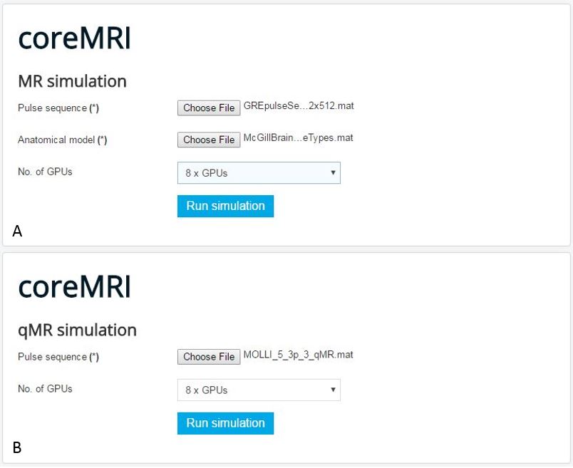
The simulation framework of coreMRI was based on a ground-up-approach design based on the principles already published in the literature4,5,6. Amazon Web Services (AWS - aws.amazon.com) were utilized for the distribution and process of data on GPU-based instances on the cloud. A dynamic web-page was developed in order to bridge the user with the cloud-based and GPU-based infrastructure and to activate the GPU-resources required for the MR simulations. Dynamic forms allowed for uploading the pulse sequence and the anatomical computer model (when required) by the user on the cloud (figure 1). Distribution of data was performed through the MATLAB single-program-multiple data (spmd) framework whereas CUDA-C was utilized for the computationally intensive simulations on the GPUs.
To evaluate the performance and scalability of coreMRI, the execution times of two experiments were recorded. In the first case, a Gradient Echo (GE) pulse sequence was applied to the McGill 3-D anatomical model of the brain7. The FOV of the GE pulse sequence was 30cm x 30cm, the k-space matrix was 512x512, the bandwidth of the receiver was 100 kHz and the static magnetic field was set to 1.5T. The pulse sequence consisted of 810497 discrete time-steps. The brain anatomical model consisted of four tissue types (white matter, grey matter, glial matter and cerebrospinal fluid) whereas the total number of tissue isochromats was 1887619. In the second case, a simulation-based quantitative MR experiment4 was performed. The simulated pulse sequence was a 5(3p)3 MOLLI8 of 724724 discrete time-steps whereas the spin population consisted of 592200 spins covering a large range of physiological combinations of native T1 and T2 values. T1 and T2 values of 700–1700 msec and 20–300 msec respectively were simulated with a T1 and T2 step of 5 mesc and 2 msec respectively.
For this study, amazon g2-type (up to 4 GPUs, NVIDIA GRID K520) and p2-type (up to 8 GPUs, NVIDIA K80) instances were utilized.
Results
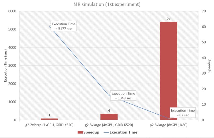
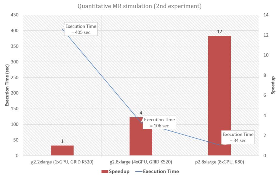
In the first experiment, coreMRI achieved with the 8 GPUs (NVIDIA K80) configuration a speedup of about 63 when compared to a single GPU (NVIDIA GRID K520) configuration (figure 2) whereas in the second experiment coreMRI achieved a speedup of about 12 (figure 3).
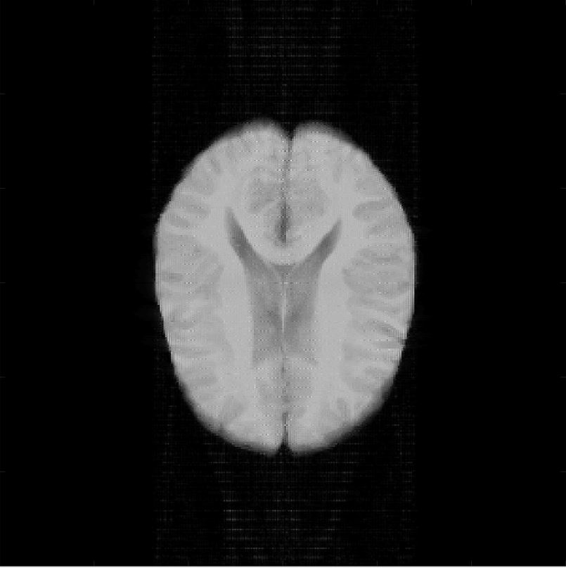
A simulated transversal tomographic image of the McGill anatomical model of the human brain is shown in Figure 4. Simulation of the entire anatomical model of the human brain, that would take more than an hour on a single-GPU computer, can now be completed within less than 2 minutes. Moreover, the development of the database of simulated MR signals to be used in quantitative MR can now be completed within clinically acceptable times (34 sec).
Conclusions
coreMRI is an online web-service available to the entire MR community. coreMRI allows its users to exploit the highly tuned computer performance of GPUs on MR simulations with neither upfront investment for purchasing advanced systems nor technical programming expertise. coreMRI is available to the users through the webpage http://www.coreMRI.com.Acknowledgements
No acknowledgement found.References
1. Benoit-Cattin, H., Collewet, G., Belaroussi, et al. The SIMRI project: a versatile and interactive MRI simulator. J Magn Reson. 2005;173:97–115.
2. Stoecker, T., Vahedipour, K. & Shah, N. J. HPC Simulation of Magnetic Resonance Imaging. Adv. Parallel Comput. 2008;15:155–164.
3. Ma, D. et al. Magnetic resonance fingerprinting. Nature 2013;495:187–192.
4. Xanthis, C. G. et al. Parallel simulations for QUAntifying RElaxation magnetic resonance constants (SQUAREMR): an example towards accurate MOLLI T1 measurements. J. Cardiovasc. Magn. Reson. 2015;17(104).
5. Xanthis, C. G., Venetis, I. E., Chalkias, et al. MRISIMUL: A GPU-based Parallel Approach to MRI Simulations . IEEE Trans. Med. Imaging 2014;3:607–617.
6. Xanthis, C. G., Venetis, I. E. & Aletras, A. H. High performance MRI simulations of motion on multi-GPU systems. J. Cardiovasc. Magn. Reson. 2014;16(48).
7. Collins, D. L. et al. Design and construction of a realistic digital brain phantom. IEEE Trans. Med. Imaging 1998;17:463–468.
8. Kellman, P. & Hansen, M. S. T1-mapping in the heart: accuracy and precision. J Cardiovasc Magn Reson. 2014;16(2).
Figures