3820
3 Dimensional Image Reconstruction – Innovation Improves Diagnosis in Pediatric MRI1Diagnostic Radiology, Phoenix Children's Hospital, Phoenix, AZ, United States
Synopsis
3D and 2D image reconstruction and other image post-processing techniques are vital in developing a complete understanding of anatomy and pathology. The ability to visualize structures in 3 dimensions, perhaps showing pathology in the orientation a surgeon will see as they approach during surgery, provide information which allows planning a procedure before the patient is under anesthesia can improve care and reduce surgical or interventional times. Further, 3D images or printed 3D models a valuable resource for teaching a patient or a child’s parent about the necessary care and treatment of their child.
Purpose
The purpose of this electronic exhibit is three-fold:
To show how 3 dimensional (3D) and image post-processing enhances diagnosis in children.
To teach the viewer how to apply image reconstruction software to pediatric disease processes.
To demonstrate methods of imaging post-processing and 3D reconstruction techniques using video to show the step by step procedures.
Outline of Content
Pediatric specific diagnoses in musculoskeletal, gynecologic, thoracic, cardiac, neurological, gastrointestinal, and vascular diseases are presented using multiple imaging modalities to correlate findings on radiographs, CT, MRI, and fusion of MRI with other modalities. Cases will be presented in a case based format including clinical information, image acquisition protocols, and will illustrate methods and diagnostic advantages of image post-processing and 3D reconstruction using a detailed video presentation format. If display space in the exhibit hall allows, 3D prints will be shown. The following cases, highlighting the following imaging diagnoses, will be presented:
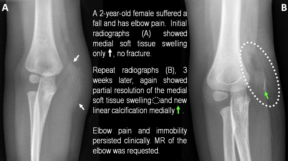
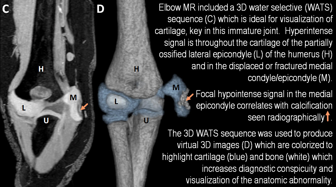
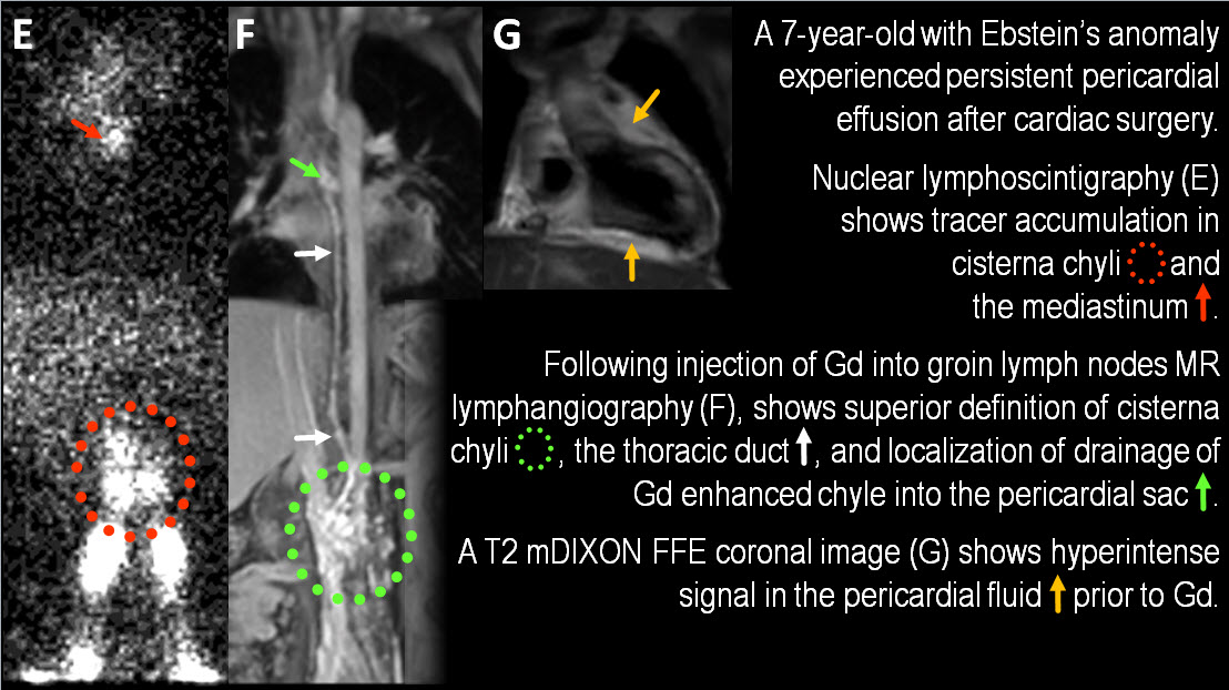
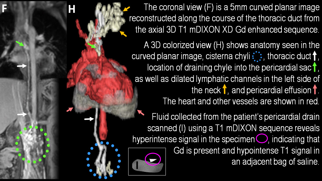
Elbow: avulsion fracture of the medial epicondyle: radiographs, MRI, 3D reconstruction Lymphangiogram: leak of lymph into pericardial sac: nuclear lymphoscintigraphy, MR lymphangiogram, 2D and 3D reconstruction
Uterine didelphys: obstructed, dilated right uterine cornu: MRI, 3D reconstruction
Pectus excavatum: MRI, 3D, lung volumes
Seizure disorder with cortical gliosis: MRI, CT, 3D reconstruction, fusion of grids on brain, WM GM volume
Cardiac/congenital heart disease: tetralolgy of Fallot: 3D volume w color definition of anatomy and chamber volume calculation
Vascular ring – airway narrowing: 3D of the trachea
Head and neck rhabdomyosarcoma: MRI tumor volume, 3D reconstruction and tracking of tumor volume through treatment course
MRCP: hepatobiliary tree anatomy: 3D reconstruction
Osteosarcoma - metal prosthesis hardware: tumor recurrence
Summary
Application of innovative 3D reconstruction and image post-processing techniques in MRI provides opportunity to enhance/improve diagnostic quality for the most accurate diagnosis and anatomic representation of pediatric anatomy and pathology.Acknowledgements
No acknowledgement found.References
No reference found.Figures