3819
IMAGING ON THE EARTH'S MAGNETIC FIELD1Division of Biomedical Imaging, University of Leeds, Leeds, United Kingdom
Synopsis
This educational abstract is aimed at providing a better understanding of NMR principles and optimisation of scan parameters for imaging using graphical illustrations and images generated with the Terranova-MRI EFNMR system. It is benefitial to clinicians who seek a more intuitive way to educate themselves with regards to MRI; to students who need hands on experience to gain a better understanding of the underlying mechanisms of signal and image formation; and to academic researchers who are looking to introduce alternative teaching methods of MRI techniques in their institutions.
Purpose
To demonstrate basic NMR principles such as signal formation, signal detection, T1 and T2 relaxation mechanisms, as well as the theory behind MR image formation and the role of MR sequence parameters on image quality and scan time, using the Terranova earth’s field nuclear magnetic resonance (EFNMR) teaching probe.Outline of Content
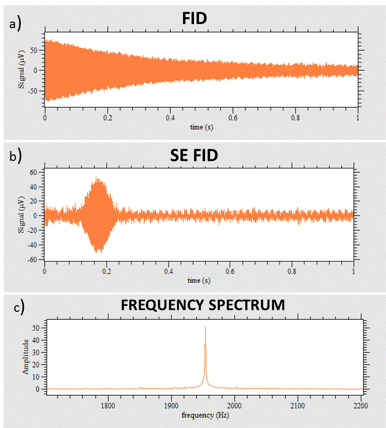
The Terranova-MRI EFNMR probe provides a cheap alternative for carrying out NMR experiments and exploring basic MRI principles1. The probe consists of a polarising coil, a B1 coil and a gradient coil1. The earth’s magnetic field is weak (50µT depending on geographical location2) and for this reason the polarising coil is used to increase the magnetisation of the sample (roughly by 350 times) prior to any excitation1,3. The net magnetisation is allowed to lie in the direction of the earth’s field before the application of a radiofrequency pulse by the B1 coil. The RF pulse applied at resonance frequency to the precessing spins, tips the net magnetisation into the transverse plane. A free induction decay is generated by the precession of the spins at larmor frequency which is detected by the B1 coil as signal1.
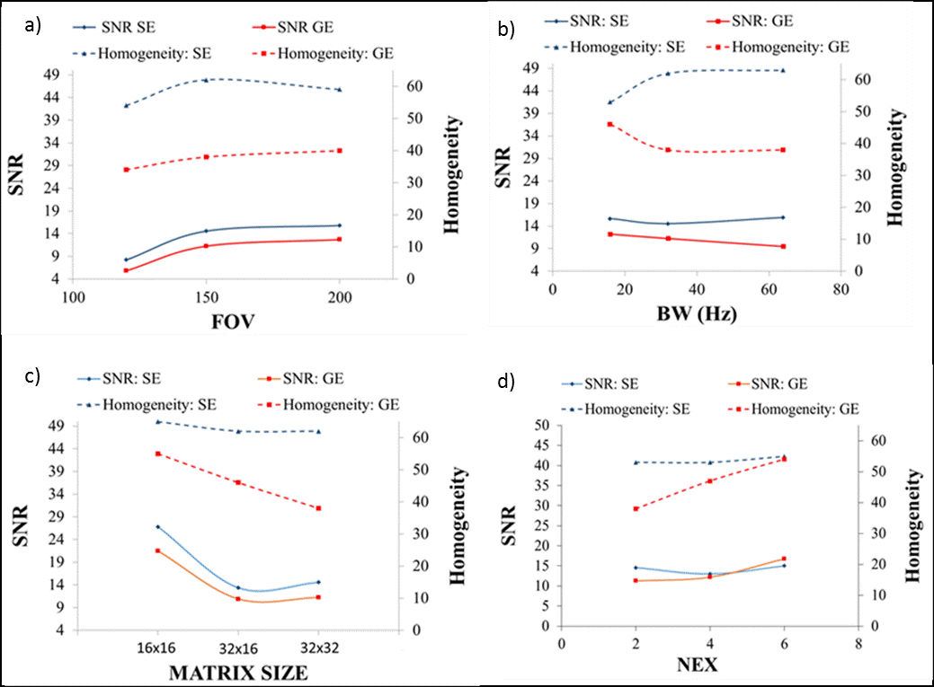
NMR measurements are highly dependent on the abundance of hydrogen molecules in a sample4. For imaging the application of gradients (through the gradient coil) control the slice thickness and the spatial encoding information received from the relaxation process of the hydrogen molecule within a particular slice. The gradient coil also shields the system from external magnetic field interference. Spin and gradient echo pulse sequences are available with the system. Spin echo (SE) imaging begins with the application of 90° pulse in the presence of a slice selection gradient. Half way through the echo time, a 180° pulse is applied to refocus the dephasing net magnetisation3,4 thus accounting for susceptibilities due to external magnetic inhomogeneities. On the other hand, gradient echo (GE) imaging sequence involves the application of a flip angle less than or equal to 90° with gradients for refocusing of the dephasing transverse magnetisation5. The concept of gradient echo imaging is to minimise scanning duration with the use of a short flip angle and refocusing gradients. However, it does not compensate for inhomogeneities in the magnetic field6. For both gradient and spin echo pulse sequences, achieving a quality image is closely linked to the optimisation of scan parameters and may lead to trade-offs between SNR, resolution and scan time7.
In this educational abstract, the Terranova-MRI EFNMR teaching system was used to:
*Illustrate the principle of spin excitation and NMR signal detection in the earth’s field (Figure 1).
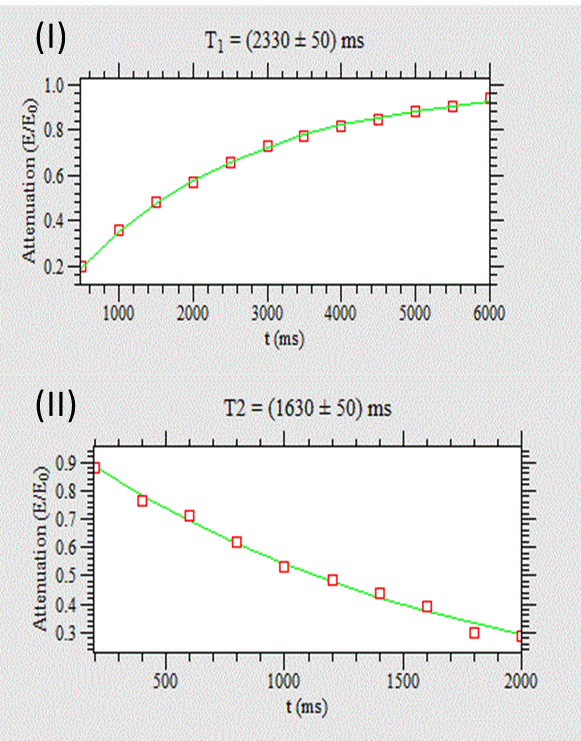
*Demonstrate the underlying theory of T1 and T2 relaxation and the basic concept of image formation (Figure 2 & 3) ·
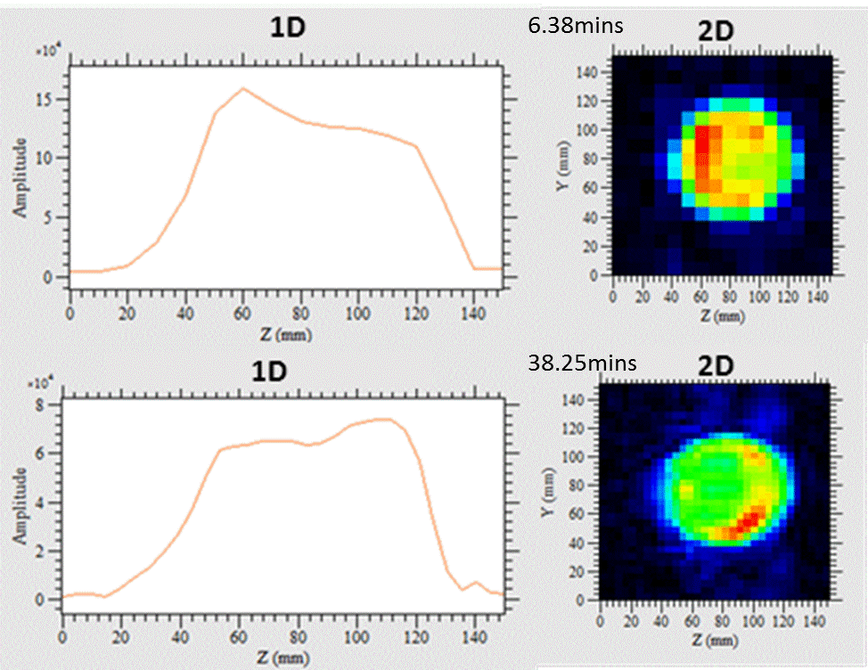
*Explain the difference between gradient echo (GE) and spin echo (SE) imaging using the earth’s field and their differences on image quality and scan time (Figure 4)
*Explore the effect of scan parameters (e.g. FOV, Matrix size, NEX and Bandwidth) on image quality and scan time (Figure 3 & 4).
Summary
The Terranova EFNRM probe provides an alternative and cheap method in better understanding basic principles governing NMR and MRI. Using graphical illustrations and images generated in the earth's magnetic field, this educational abstract unfolds complex concepts such as excitation and detection of NMR signal, as well as image formation and the effect of MRI sequence parameters on image quality and scan time.Acknowledgements
No acknowledgement found.References
1. Magritek limited. 2006. Terranova-MRI: hands on learning MRI & NMR principles, User manual. www.magritek.com
2. Mohoric, A., Planinsic, G., Kos, M., Duh, A., Stepisnik, J. 2004. Magnetic resonance imaging system based on Earth’s magnetic field. Instrumentation science and technology, 32(6), pp.655– 667.
3. Halse, M.E., Coy, A., Dykstra, R., Eccles, C., Hunter, M., Ward, R., Callaghan, P.T. 2006. A practical and flexible implementation of 3D MRI in the Earth’s magnetic field. Journal of Magnetic Resonance, 182(1), pp.75–83.
4. Hashemi, R.H., Bradley, W.G. & Lisanti, C.J. 2010. MRI: The basics. 3rd ed. Philadelphia: Lippincott Williams & Wilkins
5. Brown, M.A. & Semelka, R. 2003. MRI: basic principles and applications. 3rd ed. New Jersey: Wiley-Liss
6. Westbrook, C., Roth, C.K. & Talbot MSc, J. 2011. MRI in practice . 4th ed . Chichester: Wiley- Blackwell
7. McRobbie, D.W., Moore, E.A., Graves, M.J., Prince, M.R. 2007. MRI from picture to proton. 2nd ed. Cambridge: Cambridge university press
Figures