3810
Simulation Reveals Evidence for Bias in Parameter Estimates for Compressed Sensing of Temporally Dynamic Systems1Physics and Atmospheric Science, Dalhousie University, Halifax, NS, Canada, 2Biomedical Translational Imaging Centre, QE2 Health Sciences Centre, Halifax, NS, Canada, 3Diagnostic Radiology, Dalhousie University, Halifax, NS, Canada
Synopsis
There exists no objective framework for assessment of acquisition and reconstruction methods in compressed sensing (CS) MRI involving temporal dynamics. We propose a simulation framework to address this gap. Image quality was assessed using two quantitative metrics, and temporal parameters were recovered using least-squares fitting. CS regularization weighting was varied to determine the effect on both image quality and accuracy of recovered temporal dynamic parameters. Image quality metrics displayed distinct optima, though bias, dependent on the underlying temporal dynamics, was introduced to temporal parameter estimates. These results support the need for an objective tool to characterize CS MRI methodologies.
Introduction
Sampling rapid temporal dynamics with MRI has traditionally proven challenging, requiring trade-off between spatial and temporal resolutions. Golden angle approaches that permit retrospective choice of spatial and temporal resolutions,1 using compressed sensing (CS)2 approaches with various undersampling strategies and spatiotemporal regularizations,3 has provided further opportunity, and challenges, in the selection of this optimization. Methods for characterizing the quality of static images exist,4,5 however there is currently no framework for assessing dynamic datasets (e.g. dynamic contrast enhanced [DCE] MRI), and in particular the accuracy and precision of parameter estimates derived from the images. We propose a simulation framework to assess the effect of acquisition and reconstruction methodologies on image quality and accuracy of recovered parameters modelling temporal dynamics.
Image reconstruction quality is often quantified by the root mean squared error (RMSE), but alternative metrics from the field of image compression may better reflect the performance of CS image reconstruction. The mean structural similarity index (MSSIM) quantifies changes in structural image features,6 but has rarely been applied to CS MRI.5,7 While changes in experimental image quality can be assessed as a function of acquisition/reconstruction, without knowledge of ground truth it is impossible to determine the accuracy and precision of the parameters derived from the image sets. For initial validation we therefore simulate a phantom with temporally evolving features, and study the effect of CS regularization on image quality and recovered temporal dynamic parameters.Methods
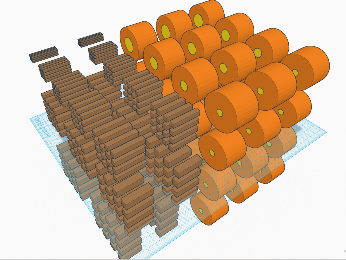
A phantom containing features with dynamically evolving signal intensity was implemented in Matlab 2016b (see Figure 1). The features allowed investigation of spatial and contrast resolutions, with temporal dynamics that broadly represent clinical scenarios (e.g. contrast agent washout). The simulated k-space was sampled using a modified CIRcular Cartesian UnderSampling (CIRCUS),8 but may be adapted for any undersampling technique (e.g. iGRASP3). Sampled CIRCUS patterns were combined within a set combination window, averaging any repeatedly sampled k-space points. CS reconstruction was performed using the Berkeley Advanced Reconstruction Toolbox.9 A total variation (TV) regularization was chosen as an exemplar because of its prevalence in the literature. The regularization weighting was varied to determine the effect on reconstructed image quality and accuracy of recovered temporal dynamic parameters. Reconstructed images were compared to the simulated image at the average acquisition time of the constituent CIRCUS patterns. Temporal dynamic parameter estimates were recovered using least-squares fitting and compared to the input parameters.Results
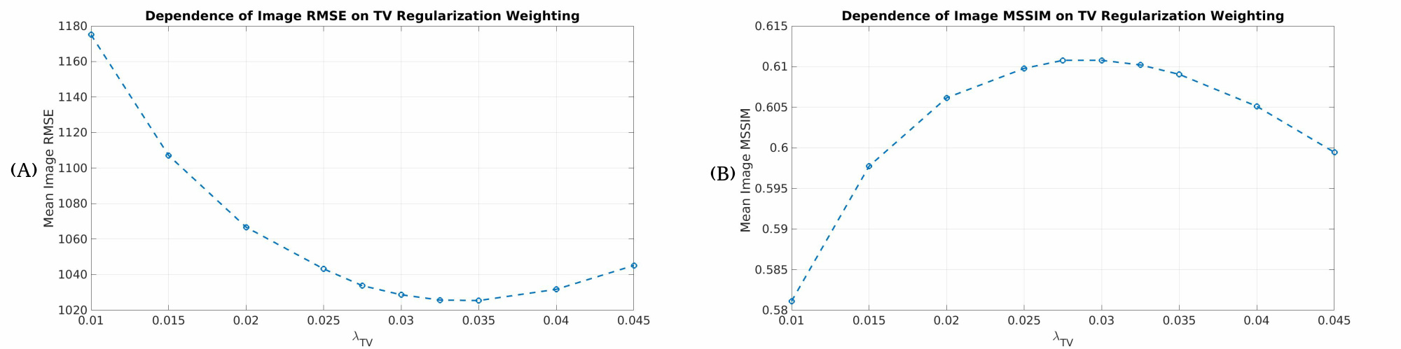
Figure 2 shows the dependence of the mean image RMSE and MSSIM on regularization weighting. A minimum in the RMSE occurred for a weighting of roughly 0.035, whereas a maximum in the MSSIM occurred for a weighting of roughly 0.03.
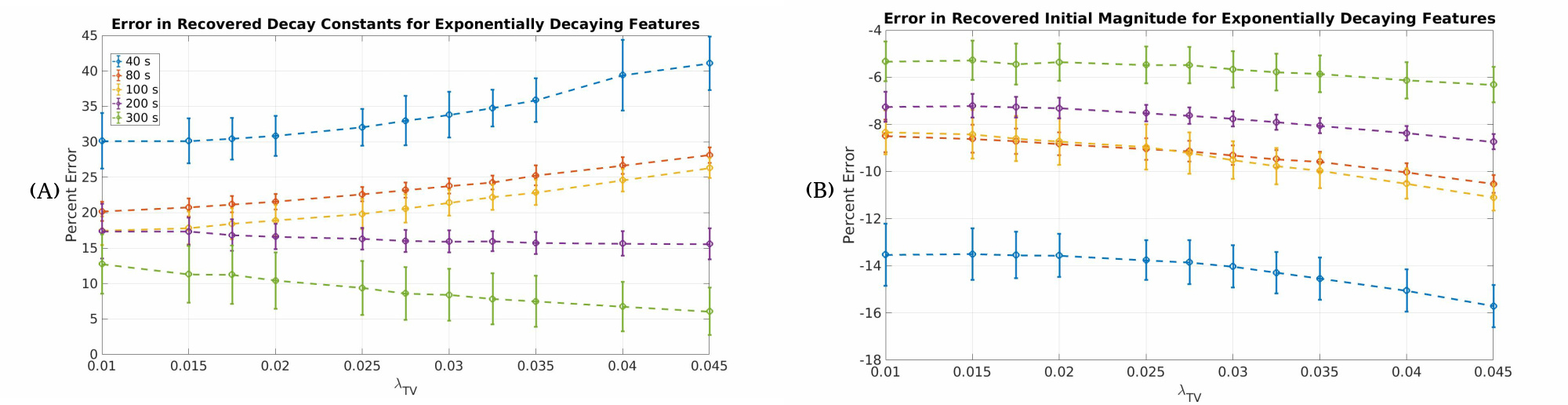
Figure 3 shows examples of the dependence of recovered temporal dynamic parameter accuracy on regularization weighting, and as a function of varying the rate of the temporal dynamics relative to the temporal resolution, for features with exponentially decaying signal intensities. The dependence between accuracy of parameter recovery and regularization weighting was dependent upon the underlying temporal dynamics.
Discussion
Figure 2 indicates that the extrema in the RMSE and MSSIM occur for different weightings, reflecting the different properties of each metric. Comparison of Figure 2 and Figure 3 suggests that high quality image reconstructions and accurately recovered temporal dynamics may require different regularization weightings. Figure 3 demonstrates that the effect of regularization weighting is dependent upon the temporal dynamics of interest. The bias in recovered parameters generally decreases as the rate of the temporal dynamics decreases relative to the fixed temporal resolution of the reconstructed images. This simulation therefore predicts a parameter estimate bias, which would be variably introduced in tissues with varying temporal dynamics, and which would go undetected in experimental data where ground truth is unknown. We have since verified this in prostate DCE data, in which retrospective resampling introduced bias in the Ktrans10 estimate that differed between healthy versus cancerous tissues (i.e. exhibiting differing DCE rates).Conclusions
Understanding of the effect of acquisition and reconstruction, with knowledge of truth, is imperative for investigating the effect of acquisition and reconstruction on temporally dynamic data. Future simulations will investigate regularizations in the temporal domain, why bias (as opposed to variability) may be introduced to recovered temporal parameters, and more. Furthermore, the simulation framework may be adapted for use with anatomically specific “pseudo-real” phantoms developed from healthy volunteer MRI data, enabling situation-specific simulations. Our initial investigations indicate the potential for data acquisition and CS reconstruction to introduce bias in parameter estimates, which could impact clinical interpretation of quantitative values such as Ktrans estimates from DCE data. This will be explored further using our simulation framework.Acknowledgements
Funding for this research was provided by the Atlantic Innovation Fund, an Investigator Sponsored Research Agreement with GE Healthcare, the NSERC Discovery Grant program (SB), and the Killam Trusts (NM).References
[1] Winkelmann S, Schaeffter T, Koehler T, et al. An Optimal Radial Profile Order Based on the Golden Ratio for Time-Resolved MRI. IEEE Transactions on Medical Imaging. 2007;26(1):68-76.
[2] Lustig M, Donoho D, Pauly J. Sparse MRI: The application of compressed sensing for rapid MR imaging. Magnetic Resonance in Medicine. 2007;58(6):1182–1195.
[3] Feng L, Grimm R, Block KT, et al. Golden-Angle Radial Sparse Parallel MRI: Combination of Compressed Sensing, Parallel Imaging, and Golden-Angle Radial Sampling for Fast and Flexible Dynamic Volumetric MRI. Magnetic Resonance in Medicine. 2014;72:707-717.
[4] Guo Y, Lebel RM, Zhu Y, et al. High-resolution whole-brain DCE-MRI using constrained reconstruction: Prospective clinical evaluation in brain tumour patients. Med Phys. 2016;43(5):2013-2023.
[5] Kim SG, Feng L, Grimm R, et al. Influence of Temporal Regularization and Radial Undersampling Factor on Compressed Sensing Reconstruction in Dynamic Contrast Enhanced MRI of the Breast. Journal of Magnetic Resonance Imaging. 2015;43(1):261-269.
[6] Wang Z, Bovik A, Sheikh H, et al. Image Quality Assessment: From Error Visibility to Structural Similarity. IEEE Transactions on Image Processing. 2004; 13:1-14.
[7] Jeromin O, Pattichis M, Calhoun VD. Optimal compressed sensing reconstructions of fMRI using 2D deterministic and stochastic sampling geometries. BioMedical Engineering OnLine. 2012;11:25.
[8] Liu J, Saloner D. Accelerated MRI with CIRcular Cartesian UnderSampling (CIRCUS): a variable density Cartesian sampling strategy for compressed sensing and parallel imaging. Quant Imaging Med Surg. 2014;4(1):57-67.
[9] Uecker M, Ong F, Tami J, et al. Berkeley Advanced Reconstruction Toolbox. Proc Intl Soc Mag Reson Med. 2015;23:2486.
[10] Tofts PS, Brix G, Buckley DL, et al. Estimating Kinetic Parameters From Dynamic Contrast-Enhanced T1-Weighted MRI of a Diffusable Tracer: Standardized Quantities and Symbols. Journal of Magnetic Resonance Imaging. 1999;10:223-232.
Figures