3786
Threonine Manganese Chelate-a New Gastrointestinal Contrast Agent for MRIYan Luo1, Hao Yu1, and Yaqi Shen1
1Radiology Department, Tongji Hospital, Tongji Medical College, Huazhong University of Science and Technology, Wuhan, People's Republic of China
Synopsis
Threonine manganese chelate is a widely used nutritional food addictive. In this pilot study, we investigated the feasibility of threonine manganese chelate as a gastrointestinal contrast agent for MRI in rat models. Our study found that threonine manganese chelate aqueous solution created dark lumen on T1w, T2w and DWI images on rat models without obvious side effect. Threonine manganese chelate might be a promising enteral contrast agent for MRI on animal models.
PURPOSE
To investigate the feasibility of threonine manganese chelate as a gastrointestinal contrast agent for MRI in rat models.INTRODUCTION
Gastrointestinal MR imaging is widely used on animal models in various studies of gastrointestinal diseases, including congenital disorders, inflammatory bowel diseases and tumors. It helps to better understand the progression of a disease and to evaluate severity and treatment response. Enteral contrast agents play an important role in gastrointestinal MR imaging. According to the signal intensity, enteral contrast agents can be classified into three categories: negative, positive and biphasic 1. Negative contrast agents reduce the signal intensity of the bowel lumen-dark lumen, make the high signal intensity lesions in the bowel wall more conspicuous. Our previous in vitro study showed that threonine manganese chelate, a nutritional food addictive, possesses favorable paramagnetic properties. This pilot study assess the feasibility of threonine manganese chelate as a negative contrast agent in rat models.METHODS
This study was approved by the Institutional Animal Care and Use Committee (IACUC). Threonine manganese chelate aqueous solutions of different concentrations (manganese concentrations ranged from 4mM to 0.15mM) were held in a custom-made phantom. Images were acquired on a 3T MR scanner (Discovery 750, GE Medical System, Milwaukee, Wis, USA) with a HD TR Knee PA coil using a T1-weighted (T1w) FSE sequence (TR 500ms, TE 6.28ms, Matrix 256*256, FOV 100*100mm) and a T2-weighted (T2w) FRFSE sequence (TR 4000ms, TE 81.7ms, Matrix 256*256, FOV 100*100mm) to determine the appropriate concentration to create dark lumen on both sequences (Fig. 1). Adult male Sprague-Dawley (SD) rats were fasted for 12 hours before MRI procedures. After anesthesia with pentobarbital (intraperitoneal injection, 30mg/kg), 1.5ml threonine manganese chelate aqueous solution was injected into the rectum. Animals were placed and immobilized in a supine position. Acquisition was performed on 3T with an 8-channel rat roil using the above sequences as well as a DW-EPI sequence (TR 2500ms, TE72.6ms, Matrix 256*256, FOV 50*100mm, b value 0,500,1000). Side effects were observed within 48 hours after MRI procedure.RESULTS
4mM threonine manganese chelate aqueous solution showed low signal intensity on both T1w and T2w sequences on 3T in vitro. After injected into rat gastrointestinal tract, it adequately distended the lumen and created dark lumen, enhanced the contrast between the intestinal wall and the lumen and improved the quality of MR images(Fig. 2). No obvious side effect was observed.DISCUSSION AND CONCLUSION
The inflammation and tumors of gastrointestinal tract usually show high signal intensity on T2w images. Negative enteral contrast agents create dark lumen and improve the tissue contrast of lesions in the bowel wall. Superparamagnetic iron oxides (SPIOs) is a negative contrast agent, however, its availability limits its use. Threonine manganese chelate is a widely used nutritional food addictive with stable chemical properties. In this pilot study, threonine manganese chelate aqueous solution created dark lumen on T1w, T2w and DWI images on rat models without obvious side effect. Threonine manganese chelate might be a promising enteral contrast agent for MRI on animal models.Acknowledgements
No acknowledgement found.References
1. Masselli G., Gualdi G. MR Imaging of the Small Bowel. Radiology. 2012 Aug;264(2):333-48.Figures
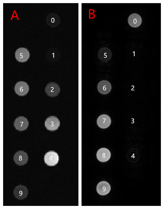
Figure 1. Threonine
manganese chelate aqueous solutions of different
concentrations on T1w and T2w sequences, (0) Distilled water, (1-9) Threonine
manganese chelate aqueous solutions, manganese concentrations ranged from 4mM
to 0.15mM, (A) Axial T1w images. (B) Axial T2w images.
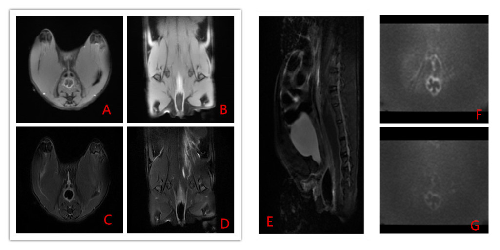
Figure 2. 4mM threonine manganese
chelate aqueous solution created dark lumen on T1w, T2w and DWI images. (A)
Axial T1w, (B) Coronal T1w, (C) Axial T2w, (D) Coronal T2w, (E) Sagittal T2w,
(F) DWI b=500, (G) DWI b=1000.