3763
Characterization of the power dependency regimes of inhomogeneous Magnetization Transfer (ihMT)1Aix Marseille Univ, CNRS, CRMBM, Marseille, France, 2Radiology, Division of MR Research, Beth Israel Deaconess Medical Center, Harvard Medical School, Boston, MA, United States, 3Aix Marseille Univ, APHM, Hôpital de La Timone, Pôle d'Imagerie Médicale, CEMEREM, Marseille, France, 4Aix Marseille Univ, APHM, Hôpital de La Timone, Pôle de Neurosciences Cliniques, Service de Neurologie, Marseille, France
Synopsis
Inhomogeneous magnetization transfer (ihMT) is a new MRI technique that shows promise for myelin imaging. A significant boost of the ihMT signal intensity is achievable when using a concentrated energy deposition scheme. Here we characterized the power dependency of ihMT for various energy deposition schemes (distributed vs. concentrated RF) and we identified distinct regimes with linear, sub-linear and saturated B1 dependency. This has a strong impact on the ihMT response to variable sequence configurations and on the most suitable conditions for different field strengths, e.g. to enhance sensitivity or provide immunity to B1+ inhomogeneities.
Introduction
Inhomogeneous magnetization transfer (ihMT) is a new MRI technique derived from classical MT that shows promise for myelin imaging [1-2]. Previous work showed that a significant boost of the ihMT signal intensity is achievable using a concentrated energy deposition scheme [3-4]. Several parameters driving this boost-effect have been investigated previously using an ihMT-GRE sequence suitable for whole brain imaging. This includes the number of consecutive saturation pulses (Np) and the repetition time (TR) between bursts of MT pulses. In the current work, special attention is paid to the RF power/B1. Experiments performed at 1.5T focused on the characteristics of ihMT vs. B1 for various energy deposition schemes spanning from distributed RF pulses (short TR) to highly concentrated bursts of RF pulses (long TR). Additionally, complementary preliminary 3T experiments assessed the sensitivity of ihMT to B1+ inhomogeneities within the whole-brain, as a function of the energy deposition schemes.Methods
Whole brain ihMT imaging was carried out using a pulsed ihMT module combined with a 3D GRE readout [3,5]. Fixed ihMT parameters were: ±Δf=7kHz, pw=0.5 ms , Δt=1ms (see definitions in Fig.1), Np=12 Hann-shaped pulses (except otherwise specified), bandwidth= 100Hz/pixel, 4.8ms TE and 2.5mm isotropic resolution. Readout segmentation and partial k-space saturation were used to reduce acquisition time, and investigate higher B1 intensities [3].
Experiment 1 was performed on a whole body 1.5-T MRI system (Avanto, Siemens, Erlangen, Germany) with a 12-channel receive-only head coil on healthy volunteers. IhMT was studied as function of the intensity of the saturation pulses, for different TR settings. Three configurations were chosen based on the characteristic of the boost effect vs TR (Fig.2c,f): 1/ TR=24ms corresponding to the most distributed energy deposition achievable with the fixed ihMT and readout parameters, 2/ TR=67ms corresponding to the optimal boosted configuration where the energy deposition was optimized to maximize the sensitivity of ihMT, 3/ TR=140ms (using higher-power Tukey pulses) corresponding to the most concentrated energy deposition considering sequence parameters and hardware limitations (peak B1). Similar experimental conditions were simulated by stepwise numerical resolution of the differential equations describing the ihMT dynamics [3] using Matlab (The MathWorks Inc., Natick, MA, USA). All results are expressed as a function of the average power defined over TR (B1rms), and the saturation power defined over the ihMT pulse train (B1sat=B1rms x √TR/(NPxΔt), Fig.1). B1rms was varied from 2uT to 5.5uT. IhMT ratios, ihMTR (fig1), were calculated in the Pyramidal Tract, PT.
Experiment 2 was performed on a whole body 3-T MRI system (Verio, Siemens) with a 32-channel receive-only head coil on healthy volunteers. IhMTR values were measured in the whole brain for B1rms = 2.2 μT and Np=6 Tukey pulses for both concentrated (TR=260ms, B1sat = 14.7 μT) and distributed energy deposition (TR=19ms, B1sat = 3.9 μT) and their dependency with the B1+ distribution was evaluated.
Results and discussion
Experiment 1: Fig.2 a-b shows experimental ihMTR as a function of B1rms and B1sat for TR=24, 67 and 140 ms. Three different patterns were found: For TR=24 ms (distributed RF power) ihMT signal slowly and almost linearly increased with B1rms over the entire range. For TR=67ms (optimized RF power), the increase was faster and tended to reach a maximum. For TR 140 ms (concentrated RF power), the ihMT signal increases even faster but rapidly saturates for B1rms > 4uT. When expressed as function of B1sat, common dependency regimes can be observed between the three configurations, with linear increase followed by strong inflexion for B1sat > 10-12uT, highlighting the dominance of saturation power (B1sat) in the ihMT effect. Simulations satisfyingly predict these observations (Fig.2 d-e).
Experiment 2: In Fig 3, the ihMTR measured in all white matter pixels is reported as a function of the local B1+ (obtained with a prepared turbo FLASH, Siemens WIP 543, and normalized to its nominal value). In addition to the ihMT boost obtained for the long TR of 260ms here, a trend for reduced B1 sensitivity was observed as compared to the distributed approach TR of 19 ms, consistent with the characteristics in B1 measured at 1.5T (Fig. 2)
Conclusion
ihMT power dependency
regimes have been studied and reveal distinct characteristics with linear,
sub-linear and saturated B1 dependency. This has a strong impact on ihMT
response to variations in sequence configurations and on the optimal conditions
for different field strengths. If low B1+ inhomogeneities are
expected, e.g. at 1.5T, a good option is to optimize TR in terms of ihMT
sensitivity (Fig.4). At 3T and higher, stronger energy concentration may be desirable
to mitigate sensitivity to B1+ non-uniformities (Fig. 3 & 5).Acknowledgements
The authors thank V. Gimenez, L. Pini ,P. Viout and E. Soulier for volunteer management. This work was supported by ARSEP foundation 2015.References
[1] G. Varma, G. Duhamel, C. de Bazelaire, and D. C. Alsop, “Magnetization Transfer from Inhomogeneously Broadened Lines: A Potential Marker for Myelin,” Magn. Reson. Med., vol. 73, no. 2, pp. 614–622, Feb. 2015.
[2] O. M. Girard, V. H. Prevost, G. Varma, P. J. Cozzone, D. C. Alsop, and G. Duhamel, “Magnetization transfer from inhomogeneously broadened lines (ihMT): Experimental optimization of saturation parameters for human brain imaging at 1.5 Tesla,” Magn. Reson. Med., vol. 73, no. 6, pp. 2111–2121, juin 2015.
[3] O. M. Girard et al., “Theoretical and Experimental Optimization of a 3D Steady-State Inhomogeneous Magnetization Transfer (ihMT) Gradient Echo Sequence: Boosting the ihMT Sensitivity with Sparse Energy Deposition,” in Proceedings of the ISMRM, Singapore, 2016, p. 2892.
[4] G. Varma, O. M. Girard, V. H. Prevost, G. Duhamel, and D. C. Alsop, “Increasing the inhomogeneous magnetization transfer (ihMT) signal in vivo with high amplitude, low duty cycle irradiation,” in Proceedings of the ISMRM, Singapore, 2016, p. 2890.
[5] O. M. Girard et al., “Whole Brain inhomogeneous MT using an ihMT prepared 3D GRE sequence at 1.5T,” in Proceedings of the ISMRM, Toronto, Canada, 2015, p. 3356.
Figures
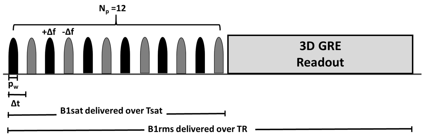
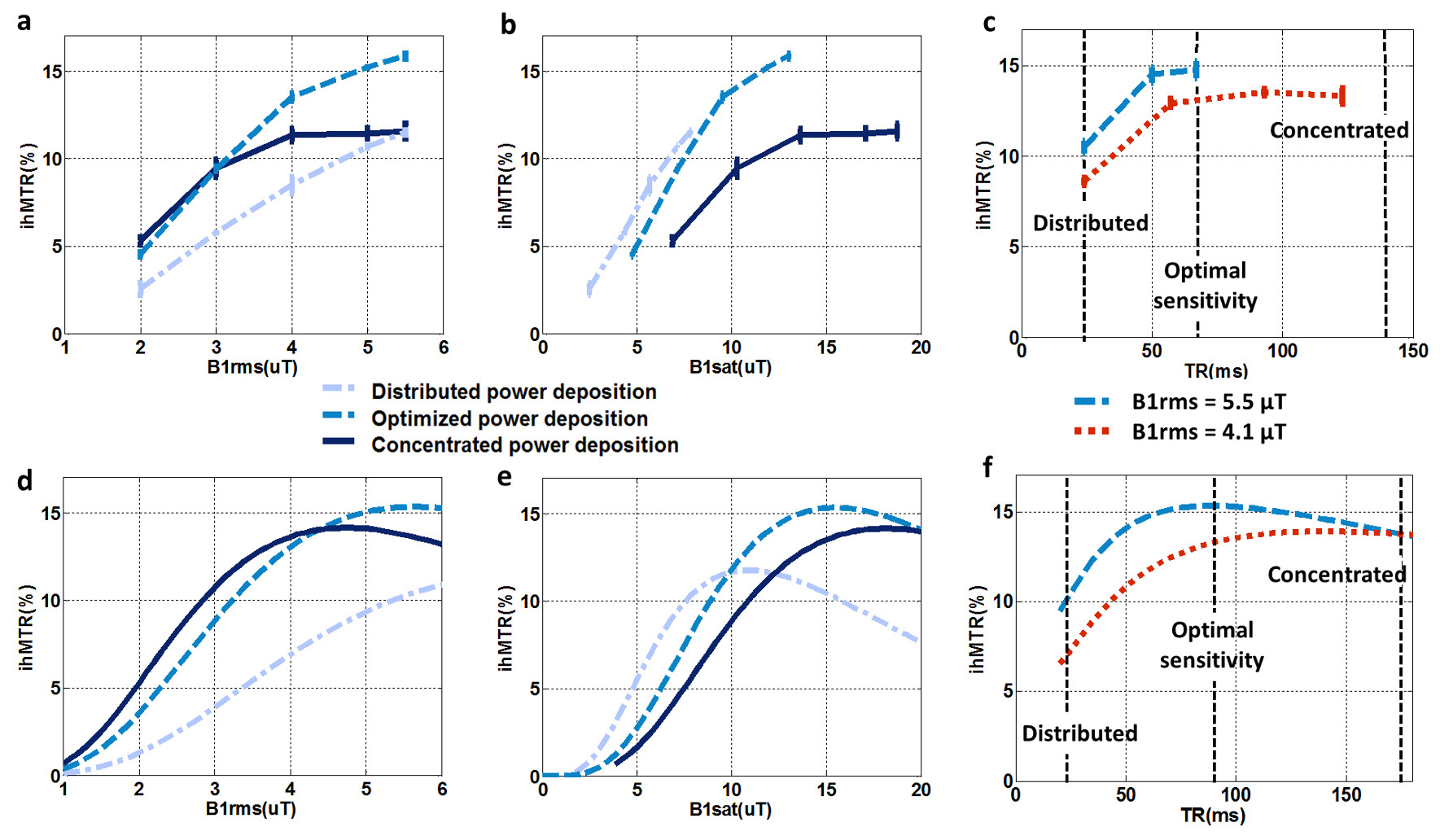
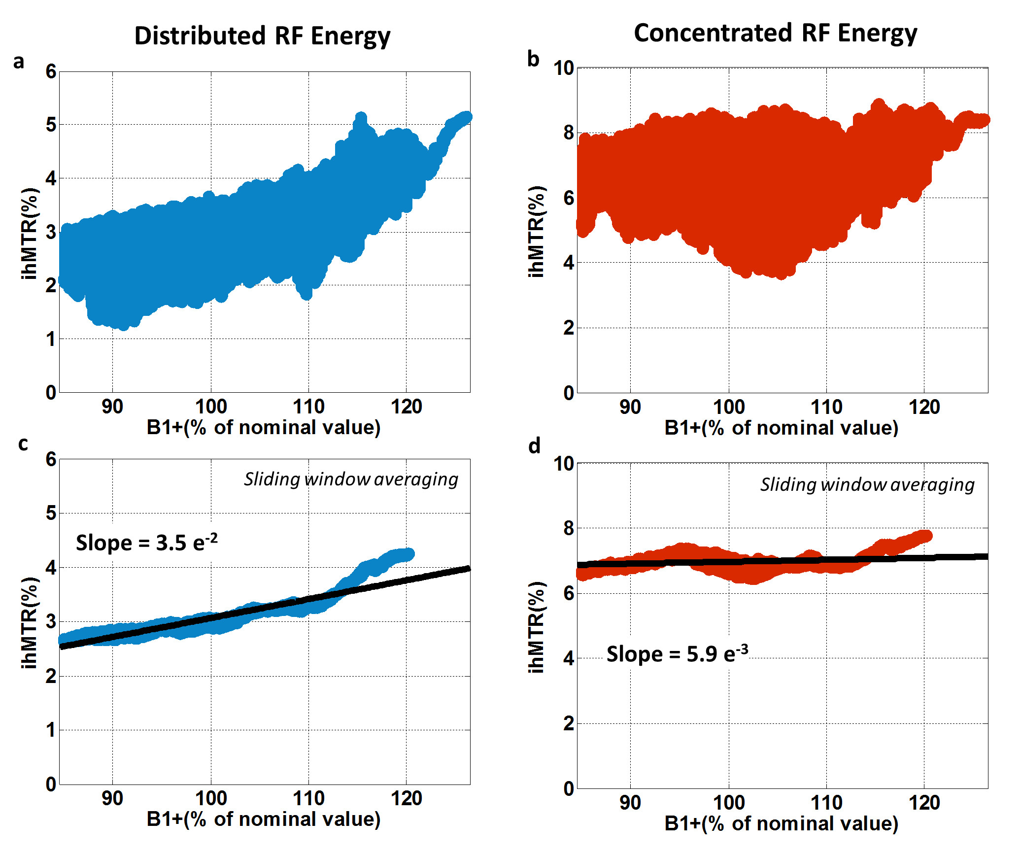
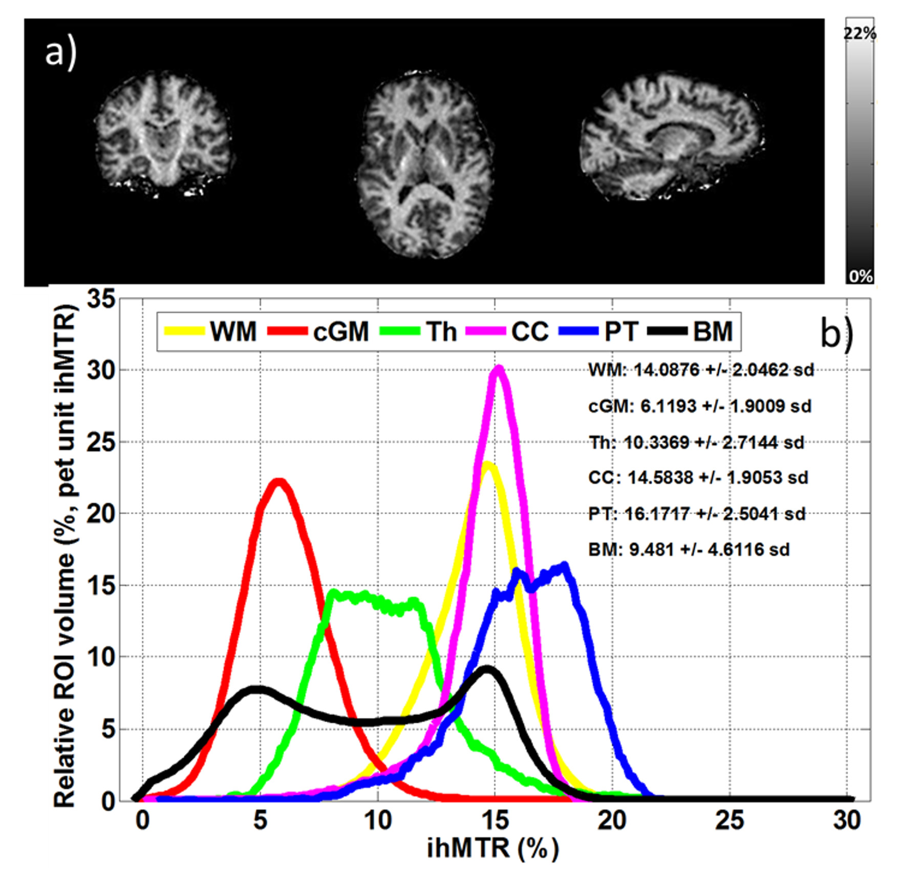
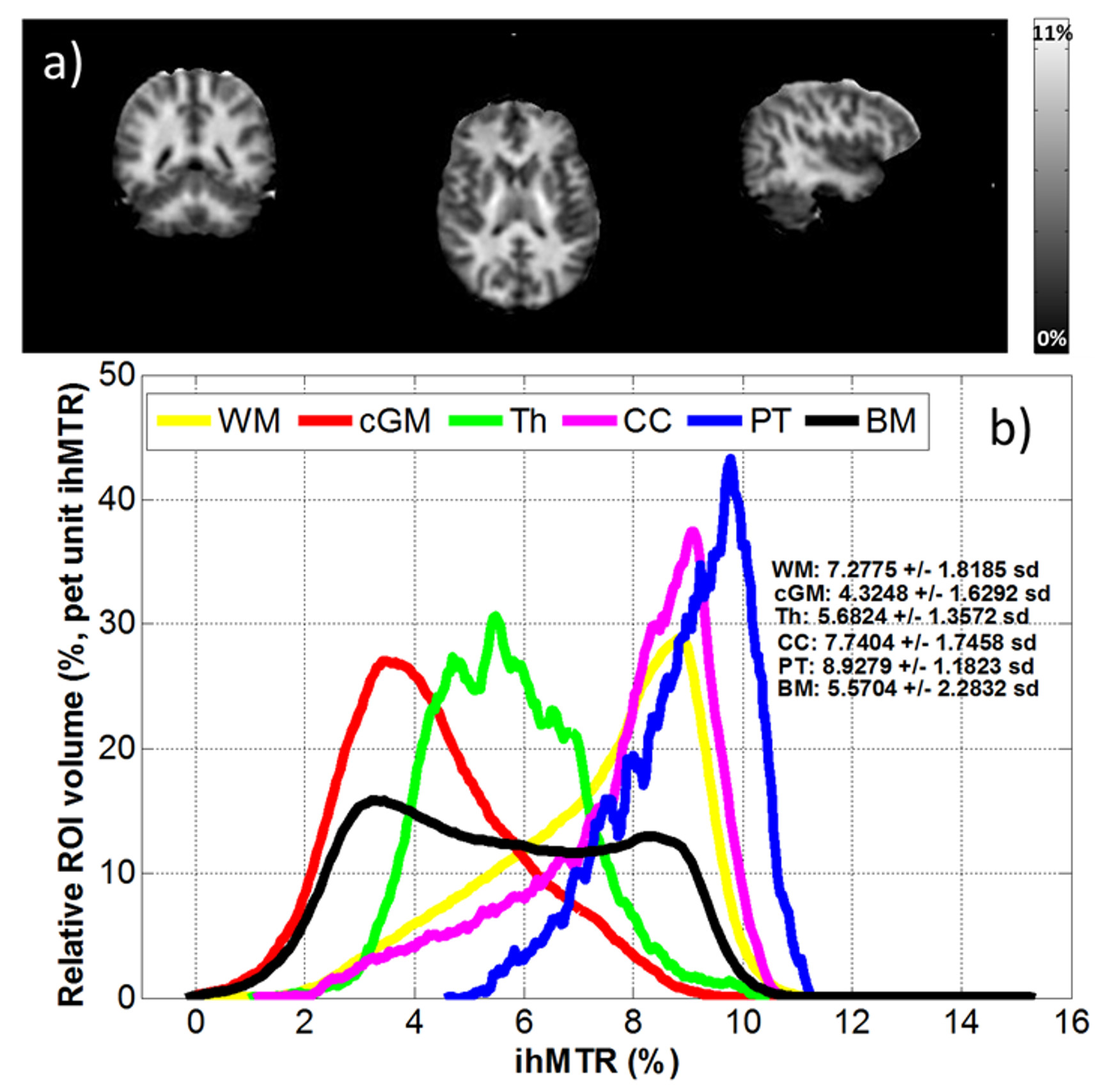
Figure 5: ihMT protocol optimized for low
sensitivity to B1+ inhomogeneities at
3T. Standard protocol: 2.5
mm isotropic, 8
min (TR=260ms,Np=6, Δf=7kHz,Tukey-pulses).
a) representative images. b) ihMTR
histograms of reference brain area (WM: whole white matter cGM:
whole cortical gray matter, Th: thamalus,
CC: corpus callosum, PT: pyramidal tract and BM: whole brain)