3718

Fast T1 mapping with temperature correction via DWI thermometry, and it application to vitreous oxygen measurement
Shengwen Deng1, Eric Muir2, Wei Zhou3, and Timothy Q. Duong2

1University of Texas Health Science Center at San Antonio, San Antonio, TX, United States, 2University of Texas Health Science Center at San Antonio, 3Radiology, Mayo Clinic

Synopsis

T1 mapping is showing great potential for mapping oxygen in human organs such as eyes and lungs. And yet, accuracy of oxygen using fast T1 imaging methods is of great concern especially in tissue with radical temperature changes. In the current study we improve fast T1 mapping with temperature correction and explore its potential in mapping 
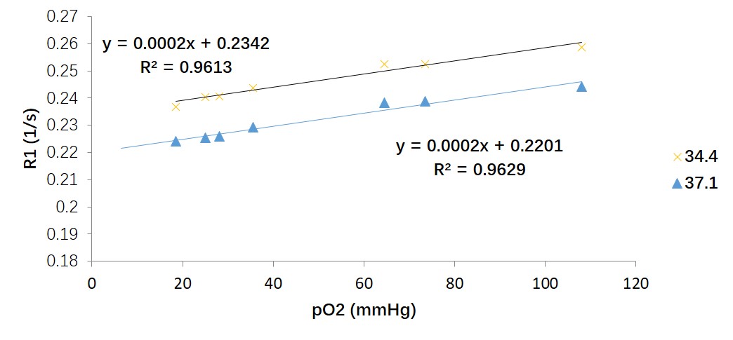
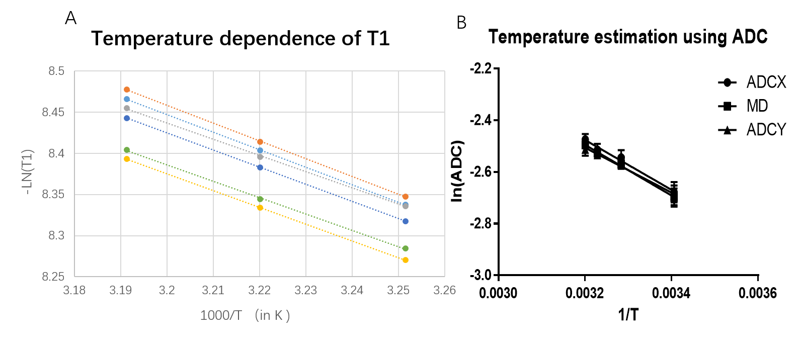
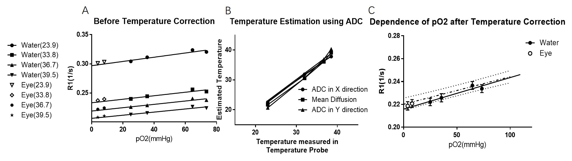
oxygen in eyes. With combination of inversion-recovery Look-Locker bSSFP and diffusion weighted thermometry, we calibrate the temperature dependence of ADC and T1, and use it to adjust the R1 for measuring partial pressure of oxygen(pO2). Fast T1 mapping could be a reliable way to pO2 that well agrees with invasive oxygen-sensitive optic fibers.

PURPOSE: Fast T1 mapping has been used to noninvasively measure partial pressure of oxygen in human vitreous due to the paramagnetic oxygen molecule. Previous studies found the accuracy of PO2 mapping is sensitive to temperature gradient across the eye between close and open condition.[1] The aim of this study is to calibrate T1 measurement with temperature correction and apply it to mapping oxygen in ex vivo eye, which is 99% water. Calibration of T1 and Apparent Diffusion Coefficient (ADC) at different PO2 and temperature were performed on water phantoms and ex vivo eyes. The temperature will be estimated via ADC-based thermometry in the phantoms, and used to adjust the T1 variation in different temperature. Non-invasive PO2 measurement with temperature correction from MRI were then compared with the exact PO2 concertation measured by oxygen-sensitive fiber optic probe. METHODS: Phantoms filled with distilled water were bubbled with nitrogen to different pO2 concentrations (n=7 for calibration and 4 for validation). pO2 was confirmed by oxygen optode (Oxylab, Oxford Optronics). Enucleated canine eyes (n=2) were kept in saline and studied within 12 hours of necropsy. After MRI, the pO2 probe was inserted into the vitreous and the phantoms to measure pO2. Samples were immersed in circulating water to control temperature from 23-40.2oC, monitored by temperature probe. MRI was performed at 3T with receive-only head coil. T1 measurements were made using inversion-recovery Look-Locker balanced steady-state free-precession (bssfp) with TE/TR=2.3/5.3ms, 21 TI from 340-10,000ms, FOV=100x100mm, matrix=176x176, 5mm slice, sequential sampling and FA=35o (and 70o for flip angle correction). Single shot Diffusion weighted EPI was used with b-value=0 and 100, TE/TR=78/6000ms. T1 and ADC were fit pixel-by-pixel. Linear regression was used to determine the slope and intercept of R1 vs pO2, ln(ADC) vs 1/Temp, and –ln(T1) vs 1/Temp (in K).[1,2]1[em1] Interclass cross correlation was used to find the agreement of estimated pO2 after temperature correction as a function of pO2 measured by invasive optode. RESULTS: With water phantoms (n=7), the relationship between R1 with pO2 at 37oC were found to be R1= [pO2]*2.00x10-4s-1/mmHg+0.2187s-1.T1 as a function of temperature with different pO2 concertation has a slope of 1.883+_ 0.08474, the R1 value of vitious is slightly higher than that of water with similar pO2. ADC as a function of temperature had slope of 8E-05/oC, with value from 0.0015 to 0.0039. ADC in different direction has different measurement error (standard error from 0.22(x-direction) to 1.68(z direction) at 36.8 oC). Using another set of phantom for validation (n=4), we found that the relation of R1 with pO2 can be merged together and giving the equation of R1= [pO2]*2.73x10-4s-1/mmHg+0.2149s-1. Ex vivo vitreous (pO2 of 8.9 and 3 mmHg from optode) had with R1 of 0.2258s-1 and 0.2235s-1 respectively at 37o, which is offset from water at 37oC and same pO2 by s-1 . Using previously measured vitreous offset of s-1 the ex vivo vitreous would have pO2 from bSSFP would be 5 mmHg and -4 mmHg[1]. Interclass cross correlation of estimated PO2 and PO2 measured after the scan is 0.9901, with a f value of 0.64 indicating good agreement between the different measurement of oxygen. DISCUSSION: The sensitivity to temperature has been considered as great impact factor in mapping oxygen in eyes. Some groups has shown that T1 could be adjusted via the linear relationship at the temperature around 35°C.[4] Our data, for the first time, quantify the temperature dependence of T1 value in fast T1 mapping methods and illustrate adjusting method that will work in physiological temperature (24 to 40°C). With this technique, the pO2 measurement in MRI can be as robust as the invasive oxygen-sensitive optic fibers in pure water and virtuous. Previously report data indicated that ADC has temperature sensitivity around 1.94-2.49%/°C[5], which agree well with our speculation. Even so, the noise and artifact in ADC maps is considered as the he major source affecting the accuracy of temperature correction. Though there has been concern that fast look-locker bSSFP mapping may underestimated the T1 value compared to inversion-recovery FLASH, our data suggests that this sequence is robust in mapping oxygen with varying range of temperature. This may provide options for imaging oxygen in eye with greater accuracy in an acceptable time. In conclusion, temperature corrected fast T1 MRI could be used to non-invasively map oxygenation of the vitreous with considerable accuracy. Future studies will improve off resonance effect in T1 mapping of in vivo eye, exploring b- value correction and eliminate the ghosting artifacts in EPI for imaging the in vivo virtuous. [em1]Hindman et al

Acknowledgements

Eric Muir, Ph.D holds a Voelcker Fund Young Investigator Award from the Max and Minnie Tomerlin Voelcker Fund.

References

Reference:

[1] Muir E R, Zhang Y, San Emeterio Nateras O, et al. Human vitreous: MR imaging of oxygen partial pressure[J]. Radiology, 2013, 266(3): 905-911.

[2] Mills R. Self-diffusion in normal and heavy water in the range 1-45. deg[J]. The Journal of Physical Chemistry, 1973, 77(5): 685-688.

[3] Hindman J C, Svirmickas A, Wood M. Relaxation processes in water. A study of the proton spin-lattice relaxation time[J]. The Journal of Chemical Physics, 1973, 59(3): 1517-1522.

[4] Simpson, Andrew RH, et al. "Measuring the Effect of Pars Plana Vitrectomy on Vitreous Oxygenation Using Magnetic Resonance ImagingEffects of PPV on pO2 Using MRI." Investigative ophthalmology & visual science 54.3 (2013): 2028-2034.

[5] Plata, Juan C., et al. "A feasibility study on monitoring the evolution of apparent diffusion coefficient decrease during thermal ablation." Medical physics 42.9 (2015): 5130-5137.

Figures

Figure 1:Relation of R1 and pO2 is effected by temperature. The temperature mostly affects the intercepts rather than the slope. All data is aquirecd using water phantoms(n=7) at 34.4 and 37.1°C.

Figure 2: Temperature dependence of T1 and ADC. Figure 2.a shows different T1 is linearly correlated with 1/Temperature (in K, unit rescaled in the figure) with R²>0.999, p< 0.0001. Figure 2.b indicate ln(ADC) (in mm2/s) is linearly correlated with 1/Temperature (in K, R²=0.9315, p< 0.0001).

Figure 3.a: Relation of pO2 vs R1 in eye and the validation phantom(n=4) are tested at various temperature, which has similar slope. 3.b: The temperature is estimated using ADC map and ADC-Temperature function derived in Figure 2. 3.c Using the estimated tempeture, the R1 value( water and eye) at various temperature are all adjusted to 37.1 and merged. The value and regression function(R²=0.9629) all fall within the 90% prediction interval of the R1-pO2 relation tested in the calibration phantom at 37.1 degree.

Figure 4: Test of agreement indicating that the pO2 value acquired via temperature corrected R1 well agrees with the value acquired via conventional method (Interclass Cross Correlation coefficient =0.9901, F value for the two measurement is 0.64, indicating the two results have good concordance.

Proc. Intl. Soc. Mag. Reson. Med. 25 (2017)
3718