3717
Motion Corrected T1 Mapping of the Pediatric Human Brain1Waisman Center, University of Wisconsin, Madison, WI, United States, 2Psychiatry, 3Medical Physics, University of Wisconsin, Madison, WI, United States
Synopsis
A method to retrospectively correct both in- and through-plane motions that occur during the acquisition of inversion recovery images is developed and used for motion-corrected T1-mapping of the pediatric human brain. Both intra- and inter-scan motions are corrected.
Target Audience
Introduction
T1 relaxometry generally requires the acquisition of several images along a parametric dimension, albeit it flip angle for variable flip angle (VFA) imaging or inversion time for inversion recovery (IR) imaging. Processing methods typically account for motion between the individual parametric images using registration methods but do nothing to handle motion that occurs during the acquisition of an individual image. While prospective methods relying on the periodic acquisition of low-resolution 2D orthogonal navigator images [1] have demonstrated promise for qualitative imaging, the effect of the extra RF pulses on the tissue magnetization in those slices is about 3-4 % and greatly complicates signal modeling due both to the variable position of the slices as well as the imperfect slice profiles created from the associated 2D excitations. In this work, we develop and demonstrate retrospective techniques to account for intra-scan motion using a 3D-radial Look-Locker acquisition termed MPnRAGE [2]. The MPnRAGE method already produces a series of IR images with different inversion times that are inherently co-registered and was previously shown to be in near perfect agreement with FSE-IR [1]. The motion correction technique does not rely on extra RF pulses or expensive camera systems and was previously demonstrated for qualitative T1-weighted imaging [3]. Here we apply similar retrospective motion correction techniques to improve the quality of quantitative T1 mapping in children.Methods
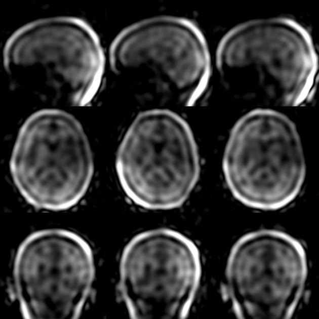
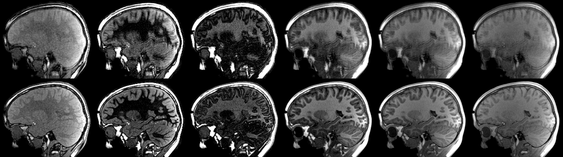
A 3D radial IR sequence, MPnRAGE, was implemented with a double bit-reversal algorithm to quasi-randomly arrange the 3D projections. The arrangement allows view-sharing both along the IR curve to reconstruct multiple images with different inversion times (TIs) as well as across/between readouts occurring after different inversion preparations. In general, MPnRAGE acquires data immediately after each inversion pulse for a period of about 1500-2000ms, which is enough data that when combined can produce a low-resolution 3D-navigator image suitable for image based registration (see Fig.1). In this manner, data collected after each inversion pulse can be co-registered. The estimated motion parameters are then applied directly to the k-space data and the different inversion time images are reconstructed.
3D-MPnRAGE was acquired on a 12 year old male with Autism using the following parameters: whole brain, non-selective excitation, 256mm isotropic FOV, 1.0mm isotropic resolution, TR/TE = 4.6ms/1.7ms, 438 points sampled along the IR curve acquired with 4 degree flip angle (points 1-326) and 6 degree flip angle (points 327-438). A 55-frame sliding-window was used to reconstruct 272 different contrast frames during the inversion recovery period. A single image with a 6 degree flip angle was reconstructed using all available data. The total scan time was 8 minutes, 33 seconds. Self-navigated retrospective motion correction was performed as described above.
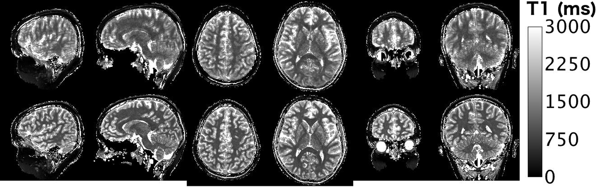
Quantitative T1 fitting was performed on a voxel-by-voxel basis similar to [2]. Briefly, an analytical formula to that accounts the two different flip angles and viewsharing was used to fit for T1, spin-density, B1 errors (for small flip angle excitations) and inversion efficiency.
Results
Please see Figures 1-3 and captions.Discussions and Conclusions
In this work, we developed and demonstrated a technique that corrects for the motion during the acquisition of the MPnRAGE data and applied this technique to minimize motion artifacts in quantitative T1 maps. The correction applies a retrospective self-navigation method and accounts for both in-plane and through-plane motions. Since all inversion times are collected for each inversion pulse, all inversion time frames are inherently co-registered. The resultant motion corrected T1 map shows much less blurring and sharper boundaries between white matter, gray matter and CSF. The quantitative T1 values in the corrected relaxometry maps are more consistent with T1 values in the absence of motion. Note that while motion can occur during the 1500-2000ms during each readout, we have previously demonstrated that that this self-navigated motion correction appears to be highly effective for correcting motion artifacts in 3D MPnRAGE T1-weighted imaging for most random and drift-type motions that are commonly observed in children [3]. Motion during each navigator image collection may result in a slight blur around the average position though this blurring appears to be minor in comparison to the larger amount of motion encountered during the entire MPnRAGE acquisition. Additional cases have been acquired are being examined to more fully evaluate MPnRAGE T1 relaxometry with retrospective motion correction.Acknowledgements
NIH grants HD079119 and HD003352 and 2RO1 MH080826-06A1.References
[1] White, N., Roddey, C., Shankaranarayanan, A., Han, E., Rettmann, D., Santos, J., Kuperman, J. and Dale, A. (2010), PROMO: Real-time prospective motion correction in MRI using image-based tracking. Magn. Reson. Med., 63: 91–105. doi:10.1002/mrm.22176
[2] Kecskemeti, S., Samsonov, A., Hurley, S. A., Dean, D. C., Field, A. and Alexander, A. L. (2016), MPnRAGE: A technique to simultaneously acquire hundreds of differently contrasted MPRAGE images with applications to quantitative T1 mapping. Magn. Reson. Med., 75: 1040–1053. doi:10.1002/mrm.25674
[3] Alexander, A, Lainhart JE, Sterling A, Travers BG, Freeman A, Kecskemeti S, (2015) Retrospective Motion Correction of MPnRAGE Studies in Children, Abstract 0833 ISMRM 2015.
Figures