3711
Development of magnetic resonance fingerprinting (MRF) combined with FISP and multi-echo SPGR acquisition for proton density, T1, T2, T2* and field mapping.1School of Electrical & Electronic Engineering, Yonsei University, Seoul, Korea, Republic of
Synopsis
Magnetic resonance fingerprinting (MRF) is a novel technique which provides rapid proton density, T1 and T2 mapping. However, susceptibility related parameters such as T2* were not acquired simultaneously. In this study, FISP and multi-echo SPGR acquisition were combined within MRF scheme to allow proton density, T1, T2, T2* and field mapping.
Purpose
Magnetic resonance fingerprinting (MRF) is a novel technique which provides rapid quantitative imaging1. In its original form, echo formalism using balanced steady state free precession (bSSFP) method was used. Two methods were recently proposed with different echo formalism, namely the fast imaging with steady state precession (FISP)2 and quick echo splitting NMR imaging technique (QUEST)3. These techniques provide quantitative information related to proton density, T1, T2 images. However, Other parameters such as T2* and field map can provide useful information. In this study, we propose a MRF technique combined with FISP and multi-echo spoiled echo gradient echo (SPGR) acquisition for proton density, T1, T2, T2* and field mapping.Method
Figure 1 shows the proposed pulse sequence diagram. FISP and multi-echo SPGR acquisitions were applied alternatively for each 200 measurements. For the FISP acquisitions, only one FISP echo was acquired for each RF excitation. However, for the multi-echo SPGR acquisitions, 30 gradient echoes were collected for each RF excitation with 50 degree of RF spoiler. For both FISP and multi-echo SPGR acquisitions, spoiler gradients (8π along the slice direction) were applied at the end of echoes. Random rotating golden angle radial acquisition5 were also applied to grant incoherency of under sampling artifacts for each measurements. A total 1000 highly under sampled (8 spokes for each images, reduction factor 32) 2D images with 3 slices were acquired in 3T Siemens scanner (Tim Trio) with 1x1mm2 resolution 256x256 mm2 FOV, 5 mm slice thickness and 391 Hz/Px bandwidth. In the FISP acquisition, echo time was set to be the half of each TR. For the multi-echo SPGR acquisitions, 1st echo time was 4.6 ms and echo spacing was 2.84 ms with collecting 30 echoes for each RF excitation. Scan time was 1 min 24 sec/slice. Each under sampled images were reconstructed using NUFFT6 with gradient delay compensation7. All experiments were performed with a healthy volunteer (IRB-approved).
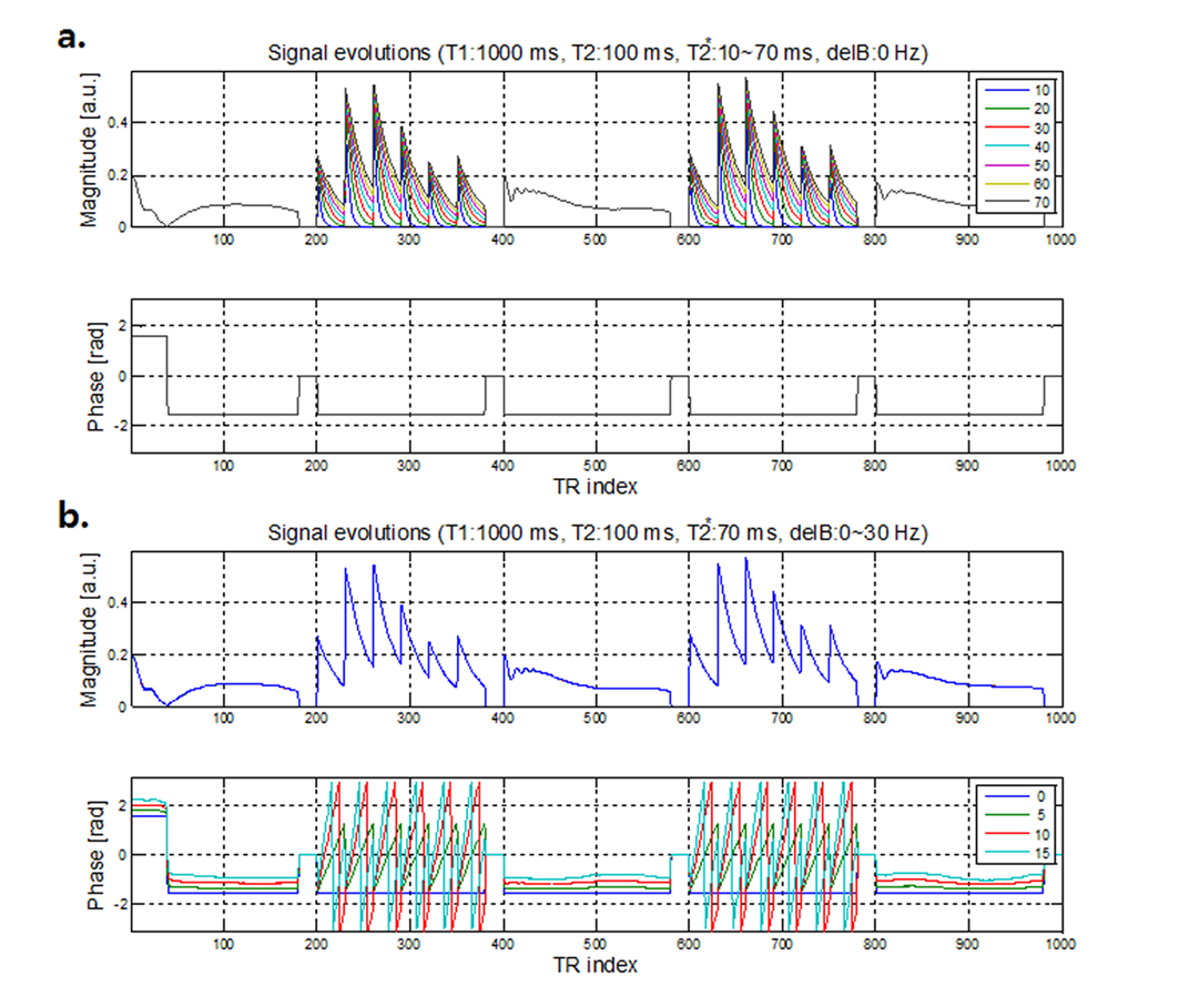
Bloch simulation was used to generate dictionary. Total 501 isochromats were calculated with different spoiling gradient along the slice direction to generate FISP signal evolution. To generate multi-echo SPGR signal evolution, T2* decay was applied during multi-echo scheme and perfect transverse magnetization spoiling was assumed after the last SPGR echo. Figure 2 shows an example of signal evolutions simulated using the proposed pulse sequence. T2* decay effect and phase variation due to the field inhomogeneity were clearly shown in Figure 2. The dictionary has T1 : [300:30:1500] ms, T2 : [21:3:150] ms, T2* : [10:2:120] ms, off resonance : [-30:5:30] Hz.
Results
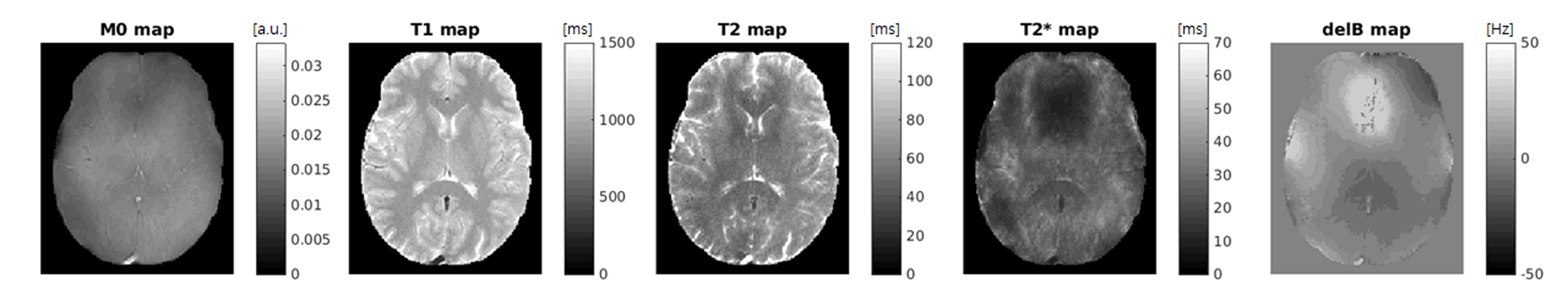
Figure 3 shows the results of the proposed method. The proposed method provides not only proton density, T1 and T2 map but also T2* and field map. T2* map shows susceptibility related contrast, such as splenium, however the image quality of T2* map requires improvement. Also B0 field inhomogeneity induced artifact are shown in T2* map, because SPGR acquisition is inherently prone to B0 field inhomogeneity.Discussion
Because of the proposed method provides various parameter information, the dictionary size became extremely huge. Therefore, matching time takes quite long (~10min) and the resolution of the one of the parameter especially off resonance parameter had a coarse step size. Further studies using MRF compression8 or Kalman filter approach9 need to be investigated The proposed method has longer scan time than spiral based MRF method, because radial acquisition was applied. Further optimization would be required to reduce the scan time.Conclusion
We have presented simultaneous proton density, T1, T2, T2* and field mapping method using a MRF technique combined FISP and multi-echo SPGR acquisition.Acknowledgements
This research was supported by Global PH.D Fellowship Program through the National Research Foundation of Korea(NRF) funded by the Ministry of Education (NRF-2015H1A2A1030698)References
1. Ma D, Gulani V, Seiberlich N, Liu K, Sunshine JL, Duerk JL, Griswold MA. Magnetic resonance fingerprinting. Nature 2013;495:187-192.
2. Jiang Y, Ma D, Seiberlich N, Gulani V, Griswold MA. MR fingerprinting using fast imaging with steady state precession (FISP) with spiral readout. Magn Reson Med 2015;74:1621-1631.
3. Jiang Y, Ma D, Jerecic R, Duerk J, Seiberlich N, Gulani V, Griswold MA. MR fingerprinting using the quick echo splitting NMR imaging technique. Magn Reson Med 2016.
4. Fujita N, Shinohara M, Tanaka H, Yutani K, Nakamura H, Murase K. Quantitative mapping of cerebral deoxyhemoglobin content using MR imaging. Neuroimage 2003;20:2071-2083.
5. Winkelmann S, Schaeffter T, Koehler T, Eggers H, Doessel O. An optimal radial profile order based on the Golden Ratio for time-resolved MRI. IEEE Trans Med Imaging 2007;26:68-76.
6. Fessler JA, Sutton BP. Nonuniform fast fourier transforms using minmax interpolation. IEEE Trans Signal Process 2003;51:560–574.
7. Block KT, Uecker M. Simple Method for Adaptive Gradient-Delay Compensation in Radial MRI. Proceedings of the 19th Annual Meeting of ISMRM, Montreal 2011;p2816.
8. Cloos MA, Knoll F, Zhao T, Block KT, Bruno M, Wiggins GC, Sodickson DK. Multiparametric imaging with heterogeneous radiofrequency fields. Nat Commun 2016;7:12445.
9. Xiaodi Zhang, Rui Li, and Xiaoping Hu. MR Fingerprinting Reconstruction with Kalman Filter. Proceedings of the 24th Annual Meeting of ISMRM, Singapore 2016;p0456
Figures
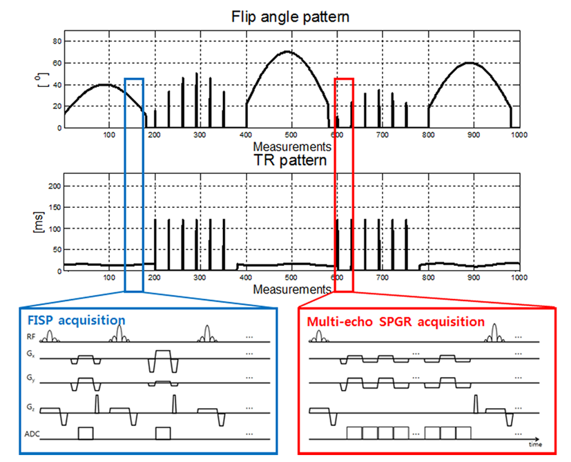
Pulse sequence diagram of the proposed method.