3706
Hyperpolarized Magnetic Resonance Imaging of Silicon Microparticles Functionalized with Mucin Antibody: Towards Molecular Targeting of Colorectal CancerNicholas Whiting1, Jingzhe Hu1,2, Julie X Liu3, Klaramari Gellci1,4, Pamela Constantinou3, Jennifer Davis5, Niki Zacharias Millward1, David G Menter6, Daniel Carson3, and Pratip Bhattacharya1
1Department of Cancer Systems Imaging, The University of Texas MD Anderson Cancer Center, Houston, TX, United States, 2Department of Bioengineering, Rice University, Houston, TX, United States, 3Department of Biosciences, Rice University, Houston, TX, United States, 4Department of Biomedical Engineering, Wayne State University, Detroit, MI, United States, 5Department of Epidemiology, The University of Texas MD Anderson Cancer Center, Houston, TX, United States, 6Department of Gastrointestinal Medical Oncology, The University of Texas MD Anderson Cancer Center, Houston, TX, United States
Synopsis
Hyperpolarized silicon nano- and microparticles hold great promise as targeted molecular imaging agents due to their overall biocompatibility and long-lasting enhanced MRI signals. We performed dynamic nuclear polarization on silicon microparticles that were functionalized with an antibody that targets Mucin overexpression in colorectal cancer. Conjugation of the antibody to the particle surface did not affect the 29Si hyperpolarization characteristics, and in vivo imaging was attained 20 minutes after particle injection into a colorectal cancer mouse model. The goal is to develop these targeted particles as a platform technology that will allow non-invasive screening of colorectal cancer using 29Si MRI.
Introduction
Silicon particles, on the nanometer to micron size scale, are potentially well-suited to function as targeted molecular imaging agents, due to their biocompatibility, biodegradability, and simple surface chemistry that is amenable to the addition of targeting moieties and therapeutic drugs [1,2]. Hyperpolarization (HP) of these particles through solid-state Dynamic Nuclear Polarization (DNP) [3] increases magnetic resonance imaging (MRI) signals by 4-5 orders of magnitude through temporary alterations to the nuclear spin distribution. This process is aided by endogenous electronic defects that are present on the particle surface, thus precluding the need for additional exogenous radical species [4]. The resulting enhanced 29Si MR signal lasts for significantly longer than other hyperpolarized agents in vivo (tens of minutes, compared to <1 minute for many other HP species) because the spin polarization is relatively protected within the core of the particle [5]. Currently, we report our efforts to develop hyperpolarized silicon microparticles for targeted molecular imaging of early stage colorectal cancer (CRC). In the United States, colorectal cancer is the second leading cause of cancer-related deaths amongst men and women (combined) [6], despite the availability of active preventative screening measures, such as the traditional colonoscopy. As such, there is a clinical need to develop advanced imaging methods for the early detection, observation of treatment efficacy, and monitoring of disease recurrence concerning colorectal cancer. In this work, we show that the hyperpolarization process is not affected by the addition of an antibody to the silicon particle surface, and the enhanced 29Si MRI signal lasts for tens of minutes after injection into a CRC mouse model.Methods
Commercially available silicon particles (average mean diameter ~2 μm) were investigated. The particles were surface functionalized with 3-aminopropyltriethoxysilane (APTES), polyethylene glycol (PEG8), and 214D4 antibody that targets the glycosylated ectodomain of human MUC1—a transmembrane Mucin protein that is overexpressed in some forms of CRC. The particles were hyperpolarized using a laboratory-constructed 29Si solid-state DNP device [7], which is located adjacent to a 7 T small animal MRI system. Initial 29Si MR signals and room temperature T1 decays were recorded spectroscopically (α = 10°) at 7 T for particles both with and without surface functionalization. Separately, functionalized particles were injected (intratumoral) into nude mice harboring a subcutaneous, human MUC1-expressing colorectal tumor (HT29-MTX-E12). Co-registered [1H:29Si] MR imaging was performed using a dual-tuned 29Si/1H Litz coil: in vivo 29Si imaging was performed with a coronal RARE sequence (α = 90°; TR/TE: 60 ms/1.8 ms; 6.4 cm FOV; 2 mm resolution), while 1H imaging is performed with a coronal RARE scan (α = 90°), TR/TE: 1927 ms/9.5 ms with a RARE factor of 8; 6.4 cm FOV (0.25 mm resolution) and 4 averages.Results
PEGylated silicon microparticles (no antibody) highlighted the colon morphology from the rectum to the cecum of a normal mouse 5 minutes after rectal administration, demonstrating the proof-of-concept for this technique (Fig. 1). Comparing bare (no surface treatments) vs. antibody-functionalized particles resulted in similar initial 29Si MRS signal and hyperpolarized decay constant (T1 ~19 minutes) for both samples (Fig. 2). Particles functionalized with the 214D4 antibody were hyperpolarized and then injected directly into a subcutaneous CRC tumor, and provided 29Si signal within the tumor 20 minutes after injection (Fig. 3).Discussion
Demonstration that the conjugation of targeting antibodies to the silicon particle surface does not affect the hyperpolarization dynamics is a critical step in developing silicon particles as a diagnostic platform for cancer medicine, and the ability to image the particles for tens of minutes following injection demonstrates an enhanced in vivo imaging window compared to other hyperpolarized contrast agents. MUC1 is an attractive target, as it is a transmembrane glycoprotein that is expressed on the cancer cell epithelium—thus the particles do not need to be internalized by the cells for effective targeting. While these are positive strides forward, the large microparticles also proved to have limited mobility, thus necessitating the administrative method of intratumoral injection. Future studies will look to translate these advances to nano-scale particles, which are expected to exhibit improved solubility and mobility—allowing for true molecular targeting. Preliminary in vitro cell-binding studies indicate that exposure to the harsh conditions of DNP do not negatively affect the targeting ability of the antibody following hyperpolarization.Conclusion
We present our most recent work developing hyperpolarized silicon microparticles for targeted molecular imaging of colorectal cancer. When fully developed, these particles are engineered to be a platform system, where different targeting agents and therapeutic drugs can be attached for advanced molecular imaging and therapeutic interventions in the clinic.Acknowledgements
This work was funded by the MDACC Odyssey Program, NCI R25T CA057730, DoD PC131680, NCI R25E CA056452, CPRIT RP150701, MDACC Colorectal Cancer Moonshot, MDACC Institutional Research Grants, MDACC Institutional Startup, U54 CA151668, Leukemia and Brain SPORE Developmental Research Awards, NCI R21 CA185536, Gulf Coast Consortium, and NCI Cancer Center Support Grant CA016672.References
[1] Park, et al. Nat. Mat. (2009); [2] Tasciotti, et al. Nat. Nano. (2008); [3] Cassidy, et al. Nat. Nano. (2013); [4] Dementyev, et al. Phys. Rev. Lett. (2008); [5] Aptekar, et al. ACS Nano (2009); [6] SEER Cancer Statistics Review (2016); [7] Whiting, et al. J. Med. Imag. (2016).Figures
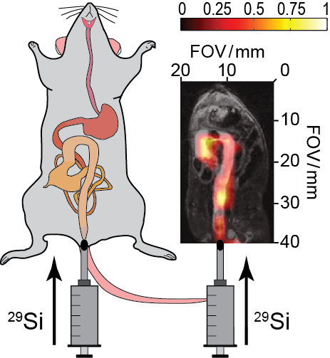
Figure 1: Proof of concept for silicon particle colonoscopy. (Left) Schematic demonstrating the rectal injection of silicon
particles into the lower intestinal tract of a normal mouse. (Right) Rectal injection of 125 mg of
PEGylated silicon microparticles (in 500 μL PBS) into a normal mouse; 29Si
image taken 5 minutes post-injection. 29Si MRI (color) overlaid on 1H MRI (greyscale) showing silicon particles occupying the intestines from
the rectum to the cecum.
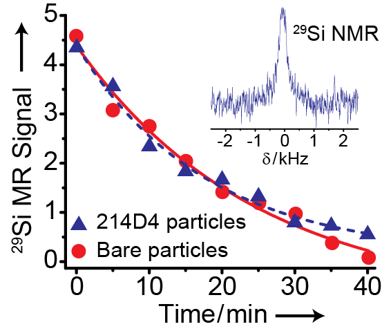
Figure 2: Hyperpolarization characteristics of targeted vs. bare particles. 29Si
MR signal decay curves for 2 μm silicon particles that are both: conjugated to
214D4 antibodies to target MUC1 (triangles),
and unfunctionalized (circles).
Samples possess similar initial 29Si signal and decay constant (T1 ~19 min for both samples).
Targeted sample consisted of 60 mg silicon particles coupled to 214D4 antibody
in 150 μL HEPES buffer (Tpol ~16.5 hrs).
Unfunctionalized sample consisted of 61 mg silicon particles in 150 μL HEPES buffer (Tpol ~18 hrs). Inset: 29Si MR spectrum from
214D4 particle sample immediately after placement into MR scanner.
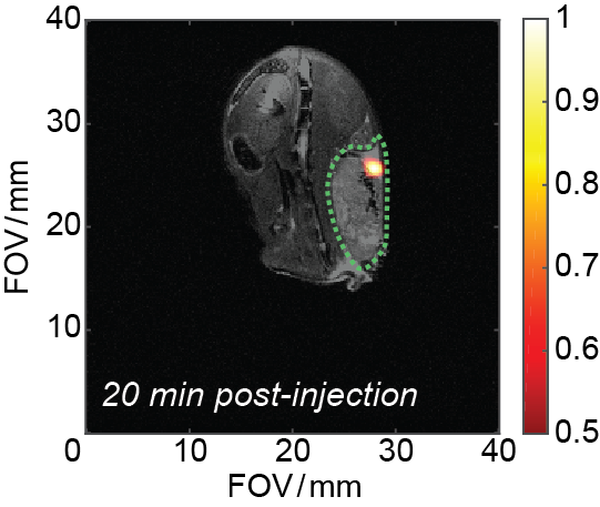
Figure 3: Long lasting HP 29Si MR signal inside tumor volume. 60 mg of
214D4-functionalized 2 μm silicon particles (in 130 μL PBS; Tpol
~18 hr) directly injected into tumor volume of a MUC1-expressing subcutaneous
HT29-MTX-E12 colorectal cancer mouse model. Image taken 20 minutes after
intratumoral injection. Silicon image (color)
overlaid with 1H anatomical image (greyscale) for co-registration; tumor volume outlined with dotted
green line.