3702
Platform for Hyperpolarized 13C MRI of Breast Cancer1Department of Medical Biophysics, University of Toronto, Toronto, ON, Canada, 2Physical Sciences, Sunnybrook Research Institute, Toronto, ON, Canada, 3LT Imaging Inc., Toronto, ON, Canada, 4Moffitt Cancer Center, Tampa, FL, United States, 5GE Healthcare, Toronto, ON, Canada
Synopsis
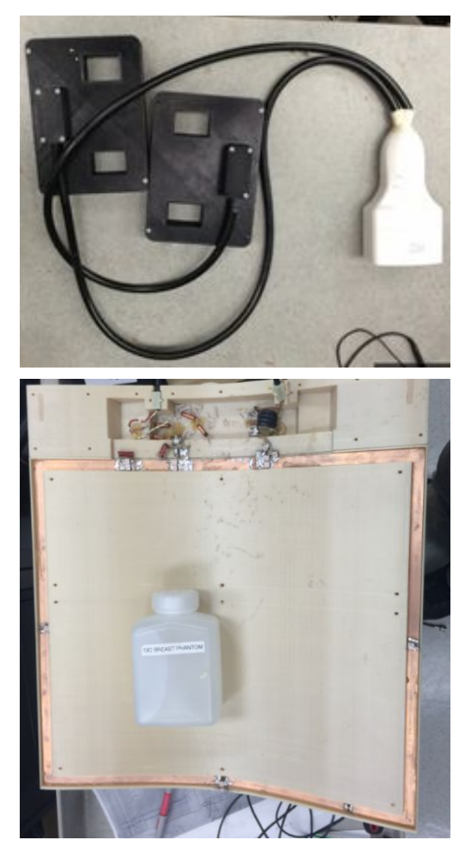
A platform consisting of a dual-echo 3D echo-planar pulse sequence and a custom coil system was implemented and tested for metabolic imaging of breast cancer. A two-channel unilateral breast 13C receive array was designed for integration with the Sentinelle Vanguard Breast MRI system. A single-loop coil was designed for RF excitation. The highly ergonomic design enables the operator to swap between proton and 13C breast coils without moving the patient, providing intrinsically registered anatomical and metabolic data. Phantom data and hyperpolarized 13C pre-clinical rat images were obtained and are presented. The proposed coil system represents important progress towards a viable breast cancer patient study.
Introduction
Pre-clinical studies of hyperpolarized 13C MRI have shown promise for visualizing abnormal metabolism associated with many cancers. Translating 13C MRI to patient studies [1,2] requires the development of tailored 13C pulse sequences and dedicated RF transmit and receive systems. In order to enable breast cancer imaging, we have designed an interchangeable coil system that allows sequential proton and 13C imaging. The design integrates with a commercially available breast imaging system, is highly ergonomic, and permits switching between proton and carbon imaging without disturbing the positioning of the patient.Methods
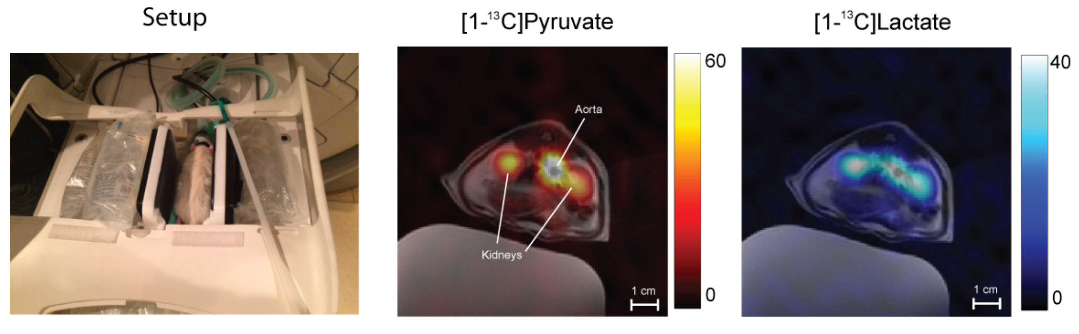
A two-channel unilateral breast 13C receive array was designed for integration with the Sentinelle Vanguard Breast MRI system (ln-Vivo Inc. Gainsville, FL) for the GE DVMR 750 3T scanner. The housing of the 13C coils are compatible with the 1H coil holders such that 1H and 13C coils can be interchanged without repositioning the patient to obtain co-registered 1H and 13C images. For 13C RF excitation, a single-loop 13C transmit coil was constructed with a 3D-printed housing that is compatible with the indentations of the removable 1H spine coil on the modular DVMR patient table such that the 13C transmitter can be secured below the Vanguard breast system. The scanner bias detunes the transmitter during 1H RF transmission to allow anatomical imaging with the 13C transmitter in place. An additional passive blocking circuit was integrated into the coil to reduce coupling with the 1H body coil, avoiding RF artefacts and power loss [3]. Imaging was performed using a GE MR750 3T scanner. The relative SNR of each receiver was assessed with a slice-selective CSI acquisition using a uniform 2.2 M 13C-urea phantom. To test the performance of the platform, a 300 g rat was placed prone between the two coil holders. A spherical 8M 13C-urea phantom was placed on top of the animal to allow 13C transmit gain calibration. Following anatomical imaging, the proton coils were removed and replaced with the 13C receivers. A 2.5 mL bolus of 80 mM pre-polarized [1-13C]pyruvate was injected via tail-vein over 12 s. An interleaved pulse sequence scheme was used, consisting of spectral-spatial excitation and 3D echo-planar imaging of lactate and pyruvate, interleaved with a slice-selective spectroscopic acquisition within each 5 s TR. The imaging was performed using a 3D dual-echo echo-planar trajectory [4] with centric phase-encoding in the slice dimension to obtain a 96×12×12 cm3 volume with nominal resolution of 7.5×7.5 mm2 in-plane and 10 mm through-plane. Signal from the two echoes were combined in magnitude to improve SNR.
Results and Discussion
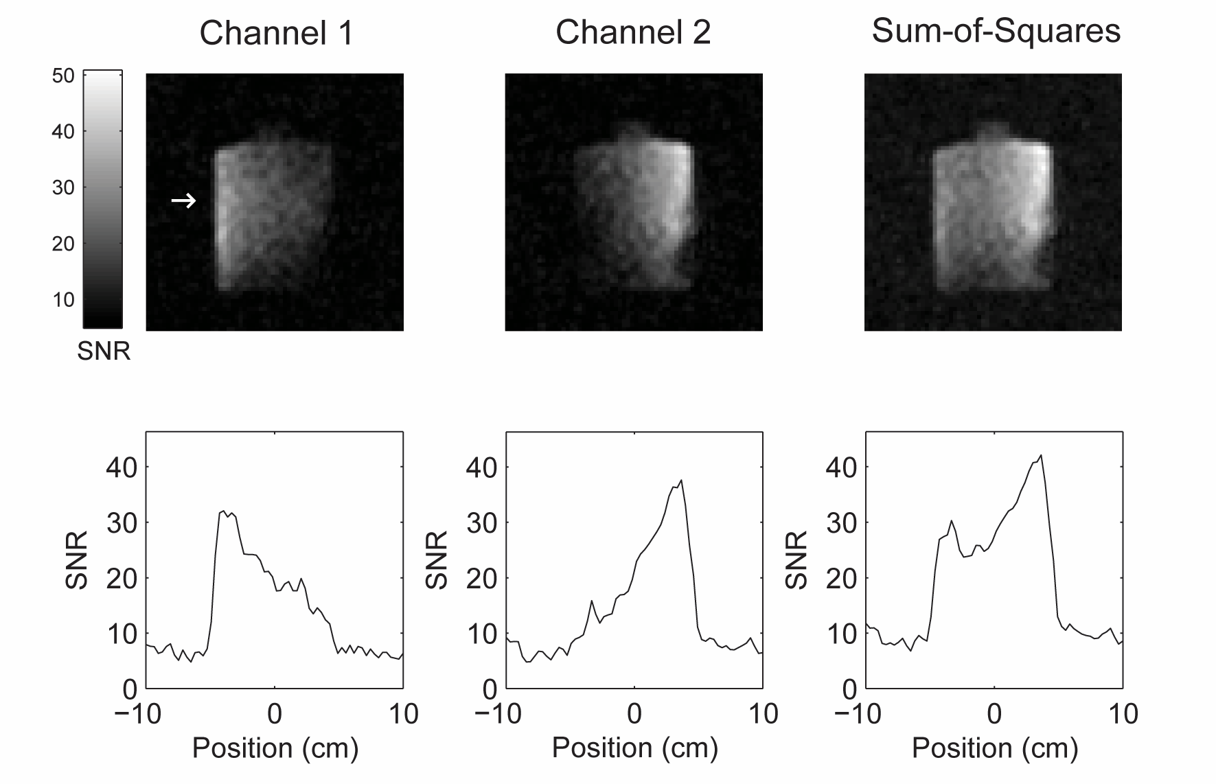
SNR vs depth profiles for each channel and the sum-of-squares combination depict the expected sensitivity profiles (figure 2). The transmit coil efficiency was measured to be 0.56 uT/√W. Time integrated hyperpolarized [1-13C]pyruvate and [1-13C]lactate images overlaid on FSE axials are shown in figure 3. A conservative choice of imaging resolution was used in these initial tests, but the resulting SNR was more than adequate and finer resolution may be achieved in future studies.Conclusion
An interchangeable coil system and tailored pulse sequence for hyperpolarized 13C MRI of breast cancer was developed and tested. Phantom experiments showed the expected sensitivity profiles and minimal coupling between channels. Metabolic imaging in a pre-clinical rat model demonstrated excellent system performance, validating the platform in advance of a human study in locally advanced breast cancer commencing shortly.Acknowledgements
The authors are grateful for funding from the Canadian Breast Cancer Foundation.References
[1] Nelson, Sarah J., et al. "Metabolic imaging of patients with prostate cancer using hyperpolarized [1-13C] pyruvate." Science translational medicine 5.198 (2013): 198ra108-198ra108.
[2] Cunningham, C.H., et al. "Hyperpolarized 13C Metabolic MRI of the Human Heart: Initial Experience." Circulation Research, 2016: CIRCRESAHA-116.
[3] Meyerspeer, M., et al., "An improved trap design for decoupling multinuclear RF coils." Magn Reson Med, 2014. 72(2): p. 584-90.
[4] Geraghty, B.J., et al., "Dual-Echo EPI Sequence for Integrated Distortion Correction in 3D Time-Resolved Hyperpolarized 13C MRI." Proceedings of the 24th Annual Meeting of ISMRM, Singapore, 2016
Figures