3632
Optimization of Flip Angle Design for Reduced T2 Blurring of 3D Arterial Spin Labeling1Radiology, Beth Israel Deaconess Medical Center and Harvard Medical School, Boston, MA, United States
Synopsis
Conventional 3D arterial spin labeling images suffer from pronounced T2 blurring. In this work, new variable flip angle schemes, which can provide a Hann or Fermi window response across the slice direction, or which can be easily corrected to the designed window response and provide optimal SNR, were evaluated. Volunteers’ results show reduced blurring, improved SNR and contrast with the proposed methods.
Purpose
Arterial spin labeling acquisition using 3D spin echo train
based sequences (e.g. stack-of-spiral [1] or GRASE [2]) is desirable because
background suppression is more optimal, the post-labeling delay is constant
across slices, and the signal-to-noise ration is high. These acquisitions
suffer, however, from blurring due to the T2 decay during the echo train. Since
the slice encoding is conventionally performed during the echo train, quite
poor slice profiles can result leading to errors such as erroneously high flow
white matter. Previous work has explored correcting T2 blurring by image
post-processing [3], reduced echo train length with
parallel imaging [4], and using a particular
variable flip angle train design [5]. Since ASL is usually a SNR
challenged acquisition, careful optimization of the acquisition SNR and point
spread function is essential. In this work, the combination of variable flip
angle trains and post processing filters were evaluated to determine optimal
SNR for prespecified point-spread-function, such as Hann and Fermi. The performance
of proposed methods was evaluated with ASL perfusion imaging on healthy volunteers. Method
Since an algorithm for simultaneous global optimization of flip angle train and filter based correction was not available, optimization was divided into two sections. First, variable flip angle trains were designed to achieve signal amplitudes, including T2 decay, matched to a number of different window functions. The extended phase map algorithm [6] was used. Echo trains were iteratively calculated as the first echo amplitude was gradually increased from zero until the target shape of response could not be achieved. Standard constant asymptotic flip angle schemes were also implemented as a benchmark. The second stage of the optimization was selection of the echo train with optimal SNR for the desired window response. In general, the optimal echo train doesn’t need to be the one that exactly produces the window response. Instead, a different echo train in combination with a correction filter may achieve a better SNR.
Five volunteers were imaged by 3D pseudo continuous ASL on a 3T GE scanner. 32 slices with slice thickness 4mm covered the majority of the brain. Five interleaved spirals with 4.1ms readout provided 4.5x4.5mm in-plane resolution for a 24cm FOV. Other parameters were labeling duration 2s, post labeling delay 1.8s, 6 repetitions, TR 4.5s, TE 9.8ms and echo spacing 9.8ms.
ASL images were processed in MATLAB. To quantify the SNR of ASL more accurately, the noise was measured by the difference from the first and last three pairs of ASL images [7]. Image quality was measured by a non-reference blurring metric [8], where large value indicated more blurred image. The contrast between grey and white matters was measured with 3D mask generated by segmenting the T1 weighted images with a probability map in SPM12. Nonparametric Friedman's test was used to compare each method with the standard flip angle scheme without correction.
Result and Discussion
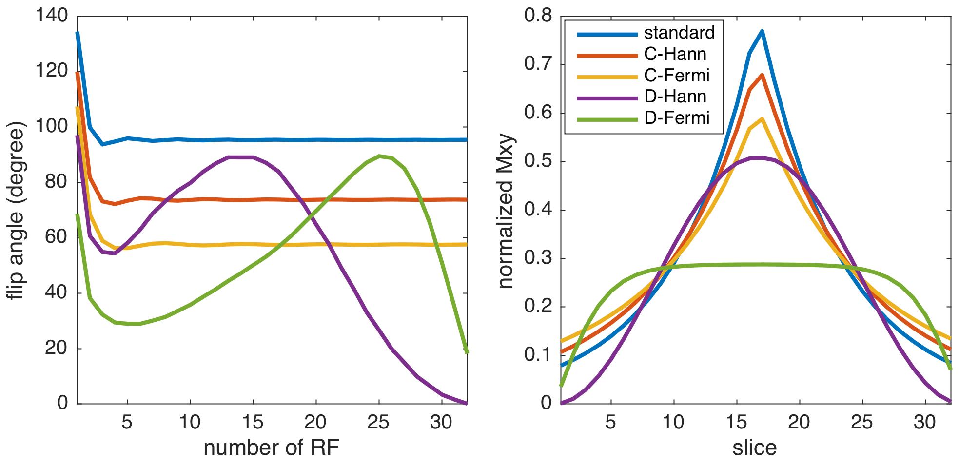
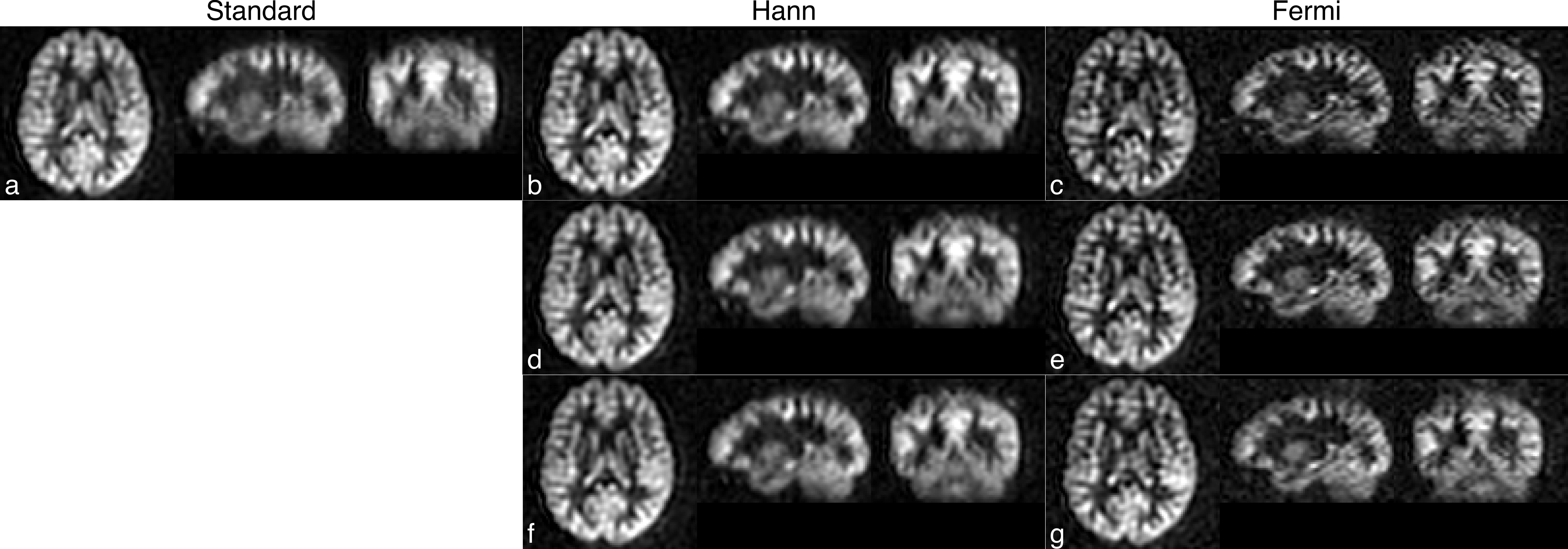
The flip angle designs and resulted echo signal are shown in Figure 1. Variable trains and filter corrections were able to achieve clear improvements in image resolution. Figure 2 shows results from one selected volunteer. With optimal flip angle and filter correction, the sharpness of ASL images was improved at the expense of SNR.
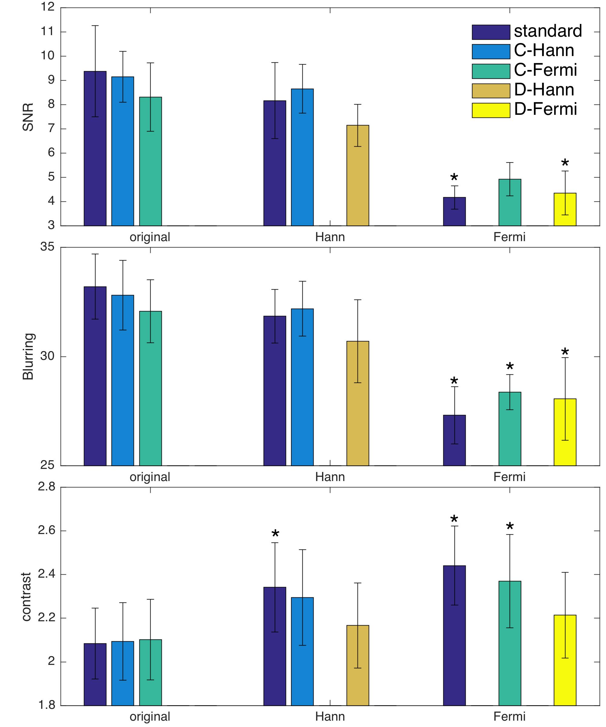
Quantitative results, Figure 3, show fairly equivalent
performance for different optimization strategies: (1) The original standard constant asymptotic flip
angle method resulted in the highest SNR (9.3±1.9), but also the most blurred images (33.2±1.5) and the lowest
contrast (2.1±0.2).
(2) When corrected to Hann window, the standard sequence resulted in significantly
higher contrast (2.3±0.2 p<0.05). The optimized
constant asymptotic flip angle for Hann windows provided the highest SNR as expected. The sequence designed for direct Hann response provided the least blurred images. (3) When corrected to
Fermi window, the constant flip angle optimized for Fermi window provided the
highest SNR, but generally the SNR of corrected image was lower than the
original ones (the standard method with Fermi window correction 4.9±0.7 p<0.005
and direct Fermi response 4.4±0.9, p<0.01). When corrected to Fermi window, all
methods showed reduced blurring (standard 27.3±1.3 p<0.001, optimal constant for Fermi 28.4±0.8 p<0.01 and
direct Fermi 28.1±1.9 p<0.01) and standard method (2.4±0.2 p<0.05)
and the constant flip angle optimal for Fermi (2.4±0.2 p<0.05) showed improved contrast.
Conclusion
Numerical optimization of multiple spin echo acquisitions suggests a combination of flip angle modulation and filter correction provides the best SNR for a desired window response. In-vivo results confirmed the resolution and contrast enhancement of the correction methods. Control of the point spread function in 3D ASL spin echo acquisitions can be achieved with a modest SNR penalty.Acknowledgements
This work is partly supported by National Institute of Mental Health; Grant number: R01-MH080729.References
[1] W. Dai, D. Garcia, C. de Bazelaire, and D. C. Alsop, “Continuous Flow-Driven Inversion for Arterial Spin Labeling Using Pulsed Radio Frequency and Gradient Fields,” Magn. Reson. Med., vol. 60, no. 6, pp. 1488–1497, Dec. 2008.
[2] D. J. Wang, J. R. Alger, J. X. Qiao, M. Gunther, W. B. Pope, J. L. Saver, N. Salamon, and D. S. Liebeskind, “Multi-Delay Multi-Parametric Arterial Spin-Labeled Perfusion MRI in Acute Ischemic Stroke – Comparison with Dynamic Susceptibility Contrast Enhanced Perfusion Imaging,” NeuroImage Clin., vol. 3, pp. 1–7, 2013.
[3] I. B. Galazzo, M. A. Chappell, D. L. Thomas, X. Golay, P. Manganotti, and E. De Vita, “Reducing blurring artifacts in 3D-GRASE ASL by integrating new acquisition and analysis strategies,” Proc. Intl. Soc. Mag. Reson. Med., vol. 22, p. 2704, 2014.
[4] M. Vidorreta, Y. V. Chang, M. A. Fernández-Seara, and J. A. Detre, “Single-Shot Whole-Brain Background-Suppressed pCASL MRI with 1D Accelerated 3D RARE Stack-Of-Spirals Readout,” Proc. Int. Soc. Magn. Reson. Med., vol. 23, p. 269, 2015.
[5] X. Liang, A. Connelly, J. D. Tournier, and F. Calamante, “A variable flip angle-based method for reducing blurring in 3D GRASE ASL,” Phys Med Biol, vol. 59, no. 18, pp. 5559–5573, 2014.
[6] M. Weigel, “Extended phase graphs: Dephasing, RF pulses, and echoes - Pure and simple,” J. Magn. Reson. Imaging, vol. 41, no. 2, pp. 266–295, 2015.
[7] O. Dietrich, J. G. Raya, S. B. Reeder, M. F. Reiser, and S. O. Schoenberg, “Measurement of signal-to-noise ratios in MR images: Influence of multichannel coils, parallel imaging, and reconstruction filters,” J. Magn. Reson. Imaging, vol. 26, no. 2, pp. 375–385, 2007.
[8] T. Dolmiere, P. Ladret, and M. Nicolas, “The Blur Effect?: Perception and Estimation with a New No-Reference Perceptual Blur Metric Fr ´ To cite this version?: The Blur Effect?: Perception and Estimation with a New No-Reference Perceptual Blur Metric,” vol. 6492, pp. 1–11, 2008.
Figures