3628
Robust 3D pCASL perfusion imaging using a Cartesian Acquisition with Spiral Reordering (CASPR)1Bioengineering, UT Dallas, Richardson, TX, United States, 2Radiology, UT Southwestern Medical Center, Dallas, TX, United States, 3Advanced Imaging Research Center, UT Southwestern Medical Center, Dallas, TX, United States
Synopsis
Arterial spin labeling can non-invasively measure perfusion, but offers low SNR compared to contrast-enhanced perfusion techniques. A novel 3D TSE with a Cartesian Acquisition with SPiral Reordering (CASPR) was implemented and combined with pCASL in the brain and kidneys. This sampling technique samples the center of k-space early in each echo train, and was shown to provide significantly improved 3D perfusion images compared to 3D linear acquisitions, and more extensive coverage than 2D acquisitions in a similar scan time.
Introduction
Arterial spin labeling (ASL) is a perfusion imaging technique with significant clinical potential due to its ability to provide perfusion maps without the administration of exogenous contrast. ASL has been well established for brain perfusion1, and is being used increasingly to measure renal perfusion2. These images are generally acquired using multiple signal averages to account for the low SNR and the characteristic significant variations among the multiple dynamic acquisitions. To overcome these challenges, 3D imaging using spiral acquisitions was recommended on a consensus paper by the ISMRM’s ASL expert panel1, since the spiral acquisitions sample the center of k-space repeatedly. However, there are several challenges in extending spiral acquisitions outside neuro imaging, particularly to body imaging with areas of increased B0 inhomogeneities. In this study, we implemented a novel 3D TSE acquisition using Cartesian Acquisition with SPiral Reordering (CASPR)3, that can be readily applied to both brain and kidney perfusion.Methods
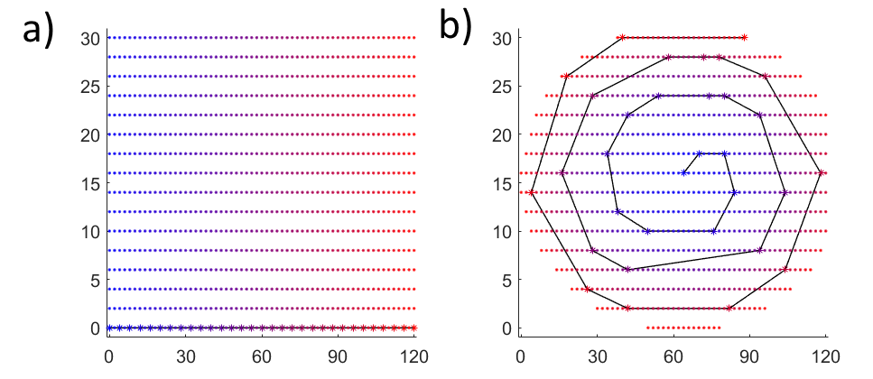
The 3D CASPR view ordering is shown in fig. 1. With this approach, each shot of the 3D TSE echo train begins by sampling the center of k-space and traversing the k-space in a spiral-out trajectory, while maintaining the sampling on a Cartesian ky-kz grid. Since the CASPR trajectory samples the center of k-space at the beginning of each echo train, this approach maximizes the prepared ASL signal allowing for longer echo trains to shorten the overall scan time, and minimize motion artifacts. The 3D CASPR view ordering3 (fig 1b) was implemented for a pCASL acquisition with background suppression and inflow saturation on a 3T Ingenia scanner (Philips Healthcare, The Netherlands). Brain perfusion images were acquired using 3D CASPR and compared against 3D TSE using a linear sampling scheme (fig. 1a), as well as multi-slice 2D gradient echo EPI in 3 volunteers. Similarly, kidney perfusion images were acquired in 3 volunteers with 3D CASPR and compared against 3D TSE with linear sampling and 2D SShTSE. The label duration and post-label delay were 1.8s for the brain1 and 1.5s for the kidneys2.
Imaging parameters for brain perfusion were:
2D EPI: scan time = 4:20, axial acquisition, 16 slices and 30 signal averages, 3x3x5 mm resolution, SENSE P reduction = 2.3
3D Linear: scan time = 4:23, sagittal acquisition, 3x3x3 mm resolution, 56 slices, halfscan factor = 0.6 along Ky, echo train length (ETL) = 86
3D CASPR: scan time = 5:34, sagittal acquisition, 3x3x3 mm resolution, 52 slices, ETL = 100
Imaging parameters for coronal kidney perfusion images were:
2D SShTSE: scan time 3:41, 3x3 mm resolution, 10mm slice thickness, 16 signal averages, halfscan factor = 0.6
3D Linear: scan time: 4:20, 3x3x3 mm resolution, 28 slices, halfscan factor = 0.7 along Ky, ETL = 90
3D CASPR: scan time: 4:20, 3x3x3 mm resolution, 28 slices, ETL = 120
Results
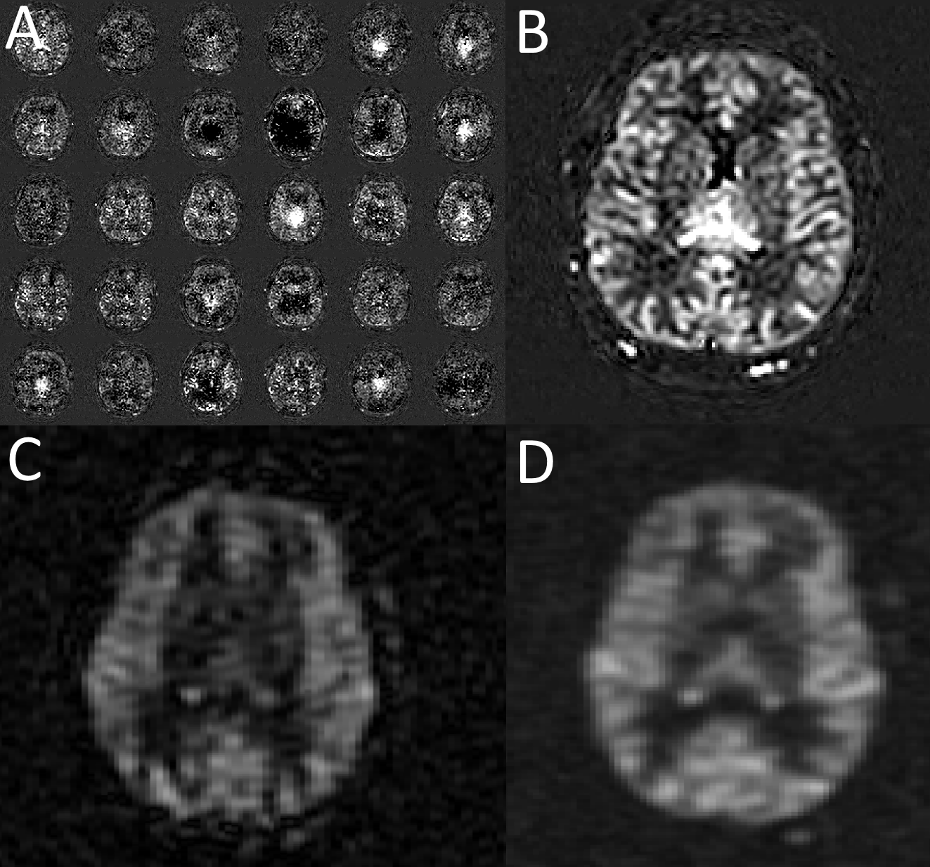
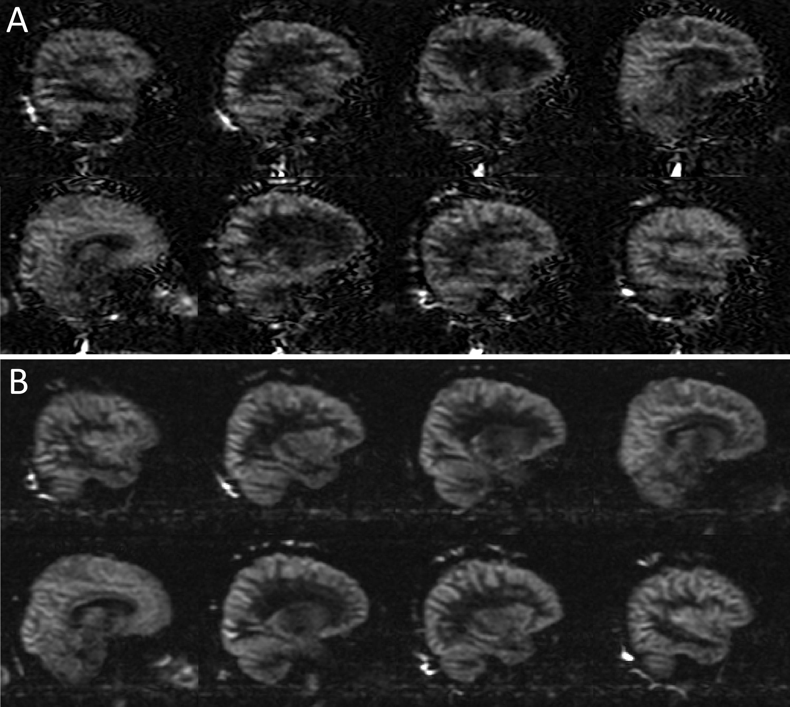
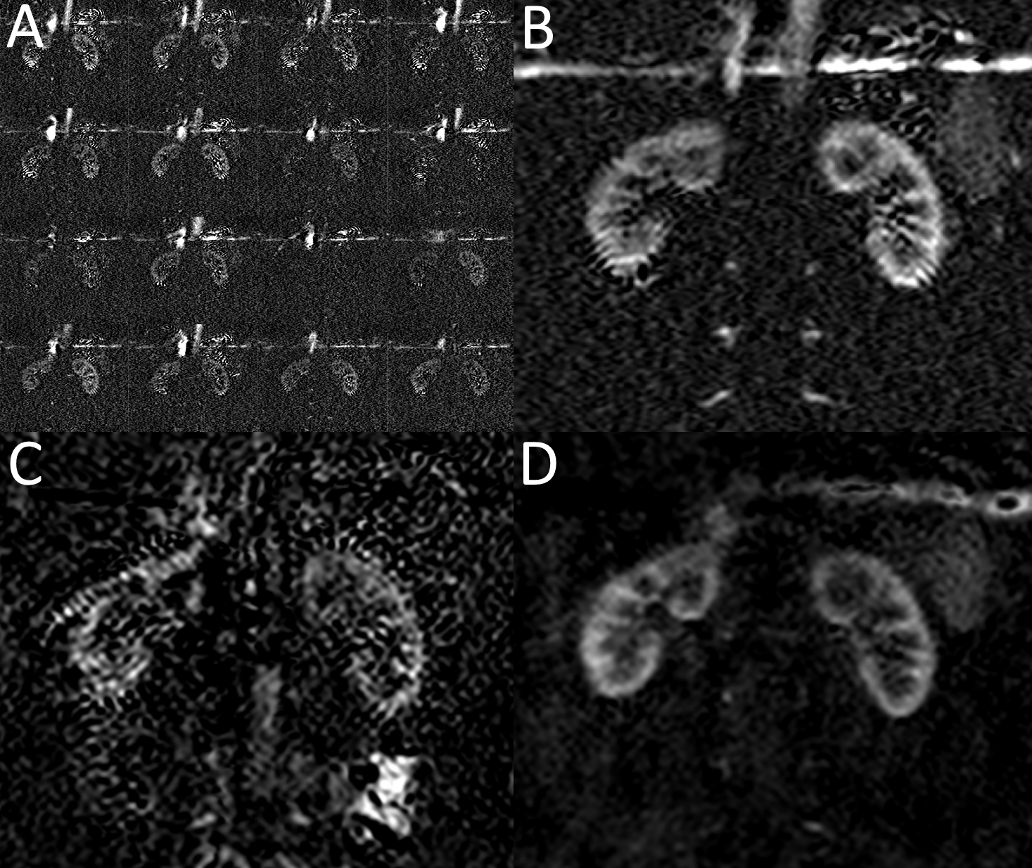
Figure 2 shows brain perfusion images using multiple repetitions of 2D EPI pCASL (fig. 2a) and the average of these repetitions (fig 2b), compared with 3D TSE acquisition using a linear (fig 2c) and 3D CASPR (fig 2d) trajectories in the same slice. 2D EPI shows significant variations from repetition to repetition and 3D CASPR shows improved image quality with higher SNR compared to 3D TSE with linear sampling. Figure 3 shows multiple slices across the brain acquired using 3D TSE with linear (fig 3a) and CASPR (fig 3b) view ordering, showing improved SNR with 3D CASPR. Similar patterns are observed in kidney perfusion (figs. 4 and 5) showing improved image quality and SNR with 3D CASPR trajectory compared against linear sampling.Discussion
With ASL acquisitions, it is critical to sample the labeled signal consistently for improved image quality and SNR. The spiral acquisitions provide this capability by repeatedly sampling the center of k-space for each repetition. Our proposed 3D CASPR trajectory provides similar capabilities, while still maintaining the sampling on a Cartesian grid and provides a more robust acquisition for 3D perfusion imaging than the linear acquisition. This also offers more complete coverage than that of 2D acquisitions in similar scan times due to improved SNR and extended echo trains. Due to the Cartesian sampling, the CASPR trajectory is readily amenable to accelerated imaging such as parallel imaging and/or compressed sensing to further reduce the scan times.Acknowledgements
No acknowledgement found.References
1. Alsop, David C., et al. "Recommended implementation of arterial spin-labeled perfusion MRI for clinical applications: A consensus of the ISMRM perfusion study group and the European consortium for ASL in dementia." Magnetic resonance in medicine 73.1 (2015): 102-116.
2. Robson, Philip M., et al. "Volumetric Arterial Spin-labeled Perfusion Imaging of the Kidneys with a Three-dimensional Fast Spin Echo Acquisition." Academic radiology 23.2 (2016): 144-154.
3. Usman, Muhammad, et. al. “Highly-efficient free breathing whole heart CINE MRI with self gated 3D CASPR-TIGER trajectory” Proceedings 24th Scientific Meeting, International Society for Magnetic Resonance in Medicine, Singapore (2016)
Figures