3624
Improved labeling efficiency in Super-Selective Pseudo-Continuous Arterial Spin Labeling1Biomedical Engineering, University of Michigan, Ann Arbor, MI, United States, 2Department of Surgery, University of Michigan, Ann Arbor, MI, United States, 3Functional MRI Laboratory, University of Michigan, Ann Arbor, MI, United States
Synopsis
We investigate how to improve the SNR of vessel-selective ASL images by off-resonance compensation and label rotation scheme optimization.
Purpose
Pseudo-Continuous
Arterial Spin Labeling (PCASL) with vessel-selective labeling has become
increasingly popular for mapping brain perfusion territories. A number of
vessel-selective labeling approaches have been proposed 1. More recently Helle et al. 2 developed
Super-selective PCASL (SSPCASL), which utilizes additional, time-varying
gradients (perpendicular to the slice-selective gradient) in between RF pulses.
Compared to standard non-selective pCASL, labeling efficiency, and consequently
SNR, are reduced in SSPCASL (see 2) The goal of this work is to
characterize and optimize SSPCASL in order to recover the full inversion
efficiency in two ways: (1) by
optimizing the rotation scheme and (2), by compensating for off-resonance
effects by adjusting the phase of the labeling pulses as in3
Methods
SSPCASL works by creating a set of labeling bands within the labeling plane by inclusion of additional gradients between RF pulses. These bands are rotated about the target vessel to define a small labeling region. The width of the labeling bands and the rotation scheme were modified to maximize labeling efficiency.
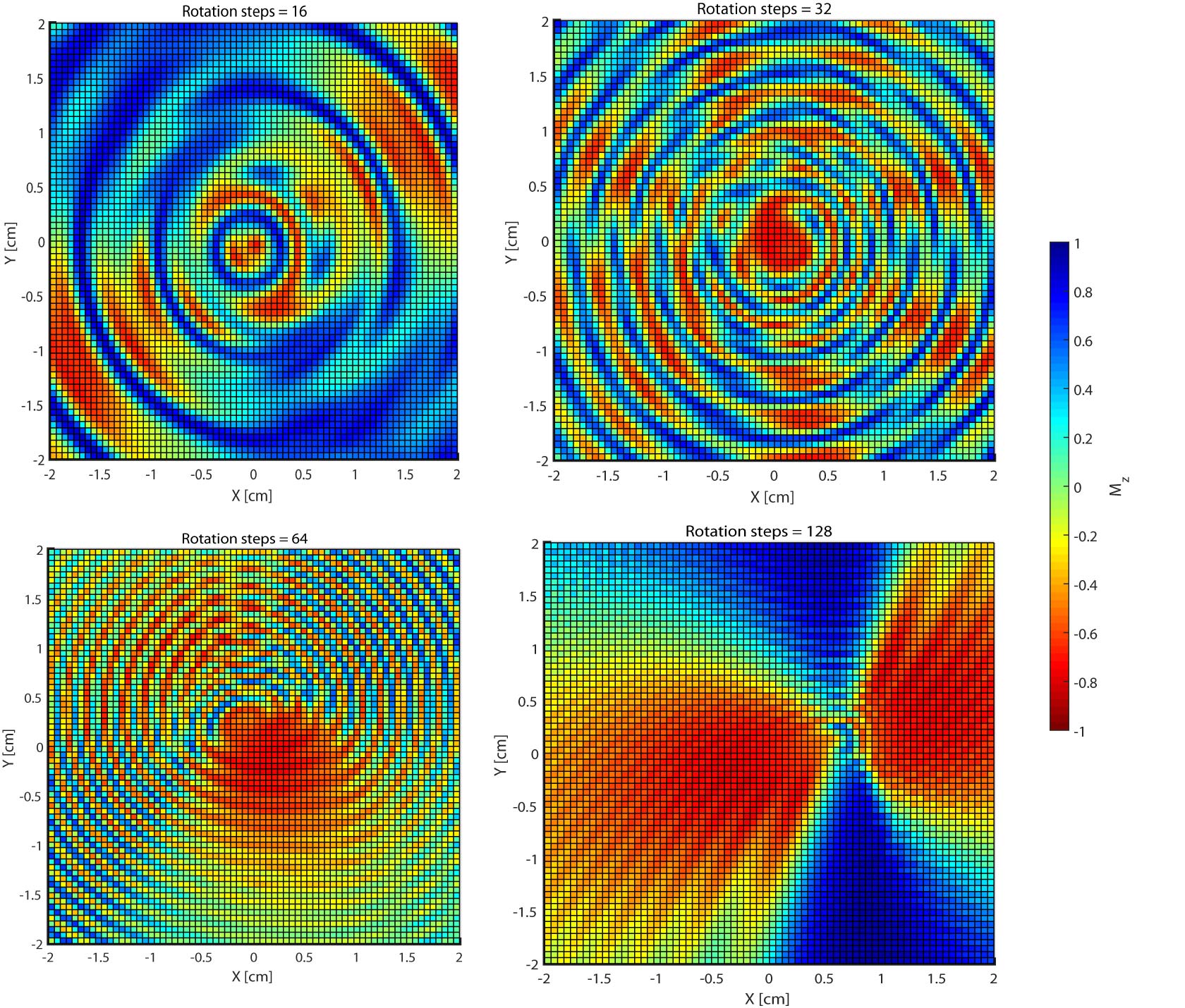
Bloch Simulations of a SSPCASL sequence were conducted to calculate a spatial map of inversion efficiency (“inversion profile”). Simulations assumed constant velocity field of 24 cm/s, 71x71 matrix, and vessel-selective gradient moment of 0.2 G/cm. Inversion profiles were compared for a range of rotation schemes.
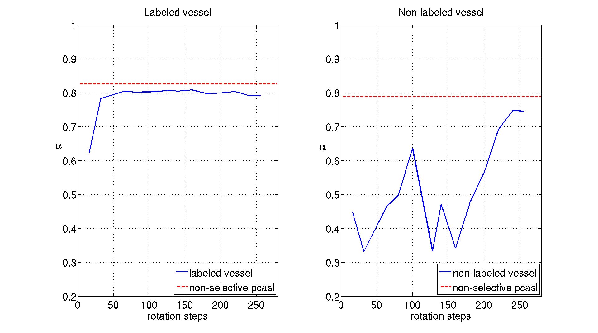
In order to validate the simulations, the following experiment was performed on a 3T MR750 scanner (GE, Waukesha,WI). A simple flow phantom consisting of tygon tubes (diameter = 6 mm), was connected to a peristaltic pump in a closed-loop . Average flow velocity was 24 cm/s. Two tubes were setup in parallel, separated by 20 mm . SSPCASL images were collected with a 2D spiral acquisition (128x128 matrix, slice thickness 7 mm , FOV 20 cm), amplitude of labeling rotation gradients = 0.22 G/cm, labeling duration = 200 ms , post-labeling delay = 50 ms. After positioning the label spot on one of the two parallel tubes, several rotation schemes were tested and compared. A 3x3 region of interest (ROI) is assigned in the center of each vessels and the degree of inversion was measured.
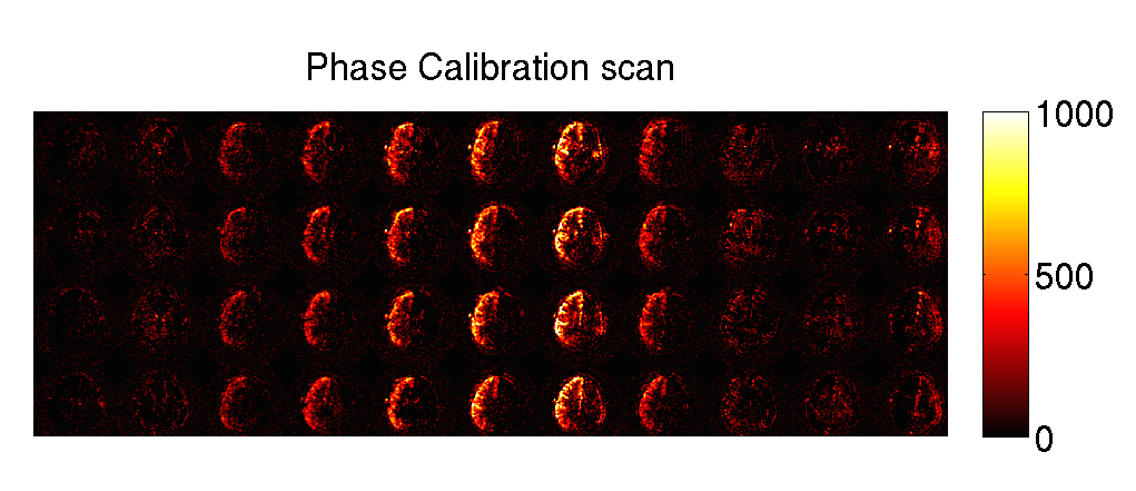
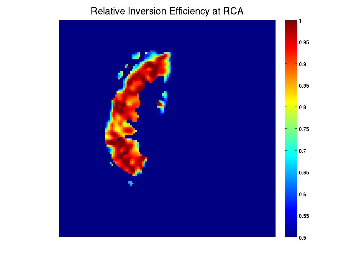
The position of left and right carotid arteries were determined from TOF images in two human participants. SSPCASL images were collected with the following scanning parameters: 3D spiral acquisition, vessel selective rotation gradient amplitude 0.24 = G/cm, number of rotations = 64, matrix size = 64x64x20 , FOV = 24 cm, slice thickness = 6 mm labeling duration = 1400 ms, post-labeling delay = 1800 ms, background-suppression pulses. In order to test for off-resonance artefacts 3, SSPCASL images were collected using a range of phase corrections (0 – PI radians) to maximize inversion efficiency empirically. Optimal phase correction was then chosen for each carotid artery and used to collect each vascular territory map. Non-selective pCASL were also collected. Inversion efficiency was calculated relative to non-selective pCASL.
Results
Figure 1 shows 4 simulated inversion profiles for different number of rotation steps using a uniform rotation scheme. The inversion efficiency in the target region (center of grid) increases with increasing number of rotation steps, but the region’s size increases also. The number of concentric circles increases with larger number of rotation steps. For 128 rotation steps, the circular shape of the label area disappears . The results of the phantom experiments (Figure 2) confirm the simulation results. With increasing number steps, inversion efficiency increased initially, and remained approximately constant for increasing number of rotation steps. In the non-labeled vessel, labeling efficiency also increased for larger number of steps. Figure 3 illustrates the results from the off-resonance calibration for the right carotid artery of one of the participants. The resulting slice images for each phase increment (column) indicate the optimal phase increment compensating for the off-resonance effects. The efficiency of SSPCASL for the right carotid artery relative to non-selective PCASL is shown in figure 4.Discussion
Simulation and experimental results showed that the inversion efficiency increases with the number of steps per rotation. However, the size of the labeling area also increases, decreasing the selectivity of the method. A smaller number of rotation steps should be chosen to optimize the selectivity profile. We are currently using the framework described here to further improve the vessel selectivity profile.
We also demonstrated in human volunteers that the off-resonance in the labeling plane can be estimated with an additional phase-calibration scan as in 3. The optimal phase correction resulted in improved labeling efficiency for SSPCASL, nearly matching the efficiency of non-selective PCASL.
Acknowledgements
No acknowledgement found.References
[1] E. C. Wong, “Vessel-encoded arterial spin-labeling using pseudocontinuous tagging,” Magn. Reson. Med., vol. 58, no. 6, pp. 1086–1091, 2007.
[2] M. Helle, D. G. Norris, S. Rüfer, K. Alfke, O. Jansen, and M. J. P. Van Osch, “Superselective pseudocontinuous arterial spin labeling,” Magn. Reson. Med., vol. 64, no. 3, pp. 777–786, 2010.
[3] H. Jahanian, D. C. Noll, and L. Hernandez-Garcia, “B0 field inhomogeneity considerations in pseudo-continuous arterial spin labeling (pCASL): effects on tagging efficiency and correction strategy,” NMR Biomed, vol. 24, no. 10, pp. 1202–1209, 2011.
Figures