3614
Metabolic characterization of rat lung transplantation using HP [1-13C]-pyruvate MRI1Radiology, University of Pennsylvania, Philadelphia, PA, United States, 2Surgery, University of Pennsylvania, Philadelphia, PA, United States
Synopsis
Orthotopic rat lung transplantation is a well-established animal model used for elucidating the mechanics of lung transplant surgery. However, most lung function assessment is conducted via invasive techniques. In this study, we demonstrated that hyperpolarized pyruvate MRI can be used to generate metabolic biomarkers that can be used for non-invasive lung function assessment after transplantation. In successful syngeneic lung transplants, the lactate-to-pyruvate ratio remains low in both lungs after transplant. However, in allogeneic or failed syngeneic lung transplantation, the native lung returns to baseline one week after surgery, whereas the transplanted lung shows a significant(~3-fold) increase in the lactate-to-pyruvate ratio.
Introduction
Orthotopic rat lung transplantation is a well-established animal model used for elucidating and improving the mechanics of lung transplant surgery, as well as recovery post-transplantation. However, most lung function assessment is conducted via invasive or post-mortem techniques [1,2]. In this preliminary study, we demonstrate the feasibility of using hyperpolarized (HP) [1-13C]-pyruvate MRI to generate metabolic biomarkers that can be used for non-invasive lung function assessment after allogeneic and syngeneic lung transplantation.Methods
Left lung allografts or isografts were transplanted from Fischer to Fischer rats (n=1, mass=250g), or Wistar Furth to Wistar Furth rats (n=2, mass~260g), respectively. A triple axis precision system was used to place and stabilize the vascular clips intrathoracically in order to clamp the bronchovascular structures, thereby avoiding interference with both the heart and contralateral lung movement. A single-suture bronchial anastomosis technique and proximal cuffing approach for vascular anastomosis was used, as previously reported [3]. The animals were monitored post-surgery to assess their recovery. HP [1-13C]-pyruvate MR imaging was performed on days 3 and 7 (days 4 and 8 for one animal). The animals were imaged in the supine position in a 4.7T magnet (Varian Inc.). HP [1-13C]-pyruvate (28.6mg, 15mM OX063, 1.5mM Dotarem Gd) was polarized using a HyperSense DNP polarizer, and ~2.5mL (8mL/kg, 80mM) of the HP agent was injected via the tail vein over a period of 6s. HP [1-13C]-pyruvate chemical shift imaging (CSI) was performed using a 2D slice selective phase-encoded FID-CSI sequence, as previously reported (TR/TE=35.7/0.35ms, α=12°, FOV=45x45x15mm3) [4]. The spectra were reconstructed, processed and analyzed using custom MATLAB scripts.Results
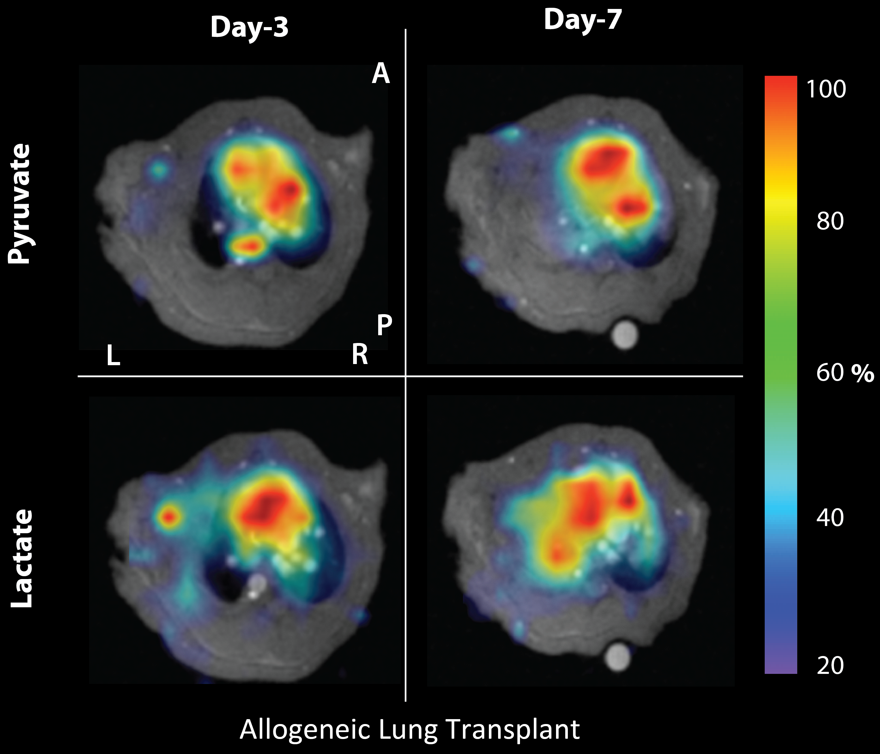
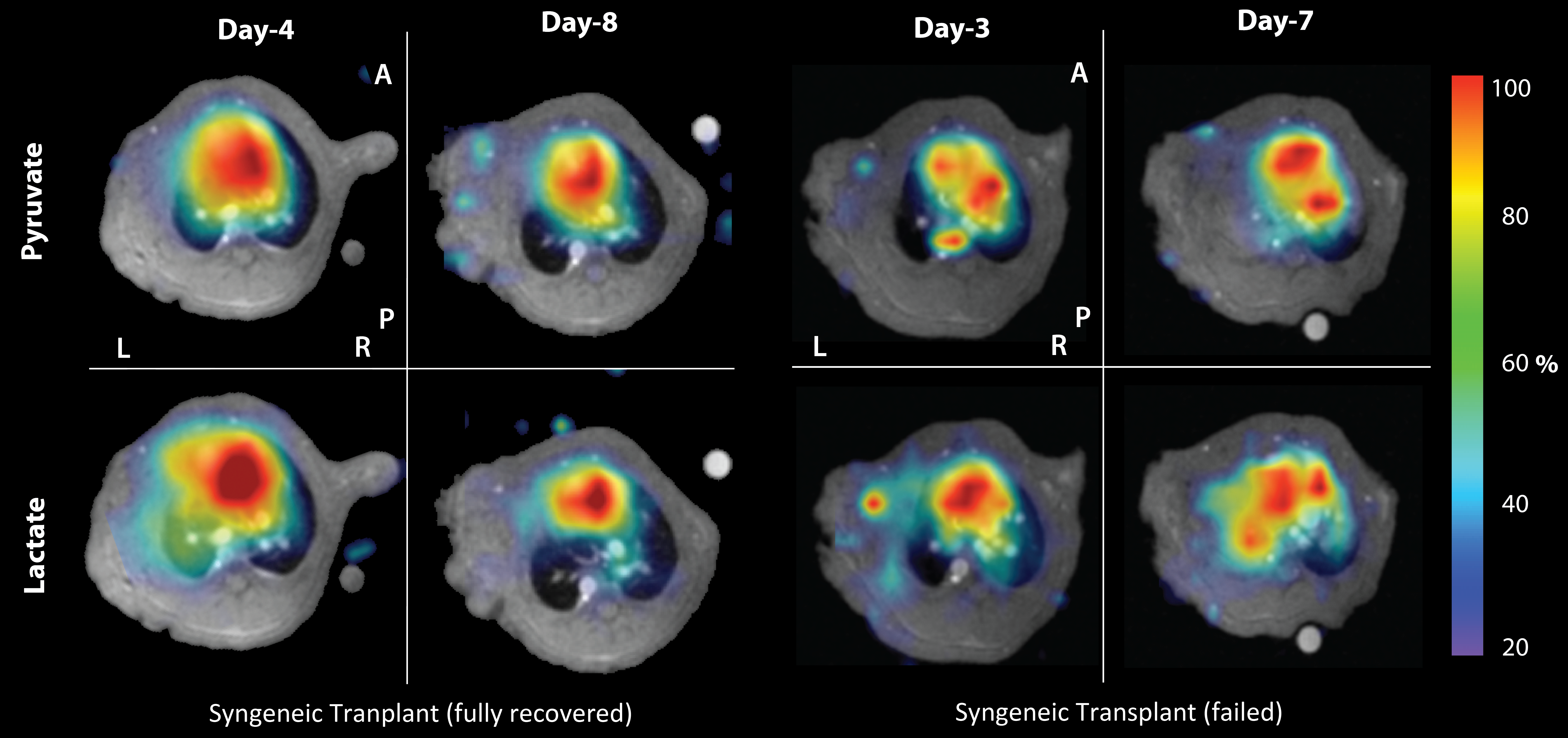
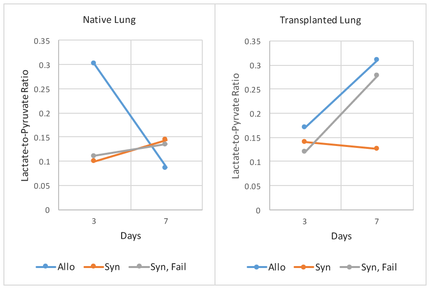
As expected, the allogeneic transplanted lung was acutely rejected (figure 1). Compared to the fully recovered syngeneic rat (figure 2, left), the pyruvate signal in the native lung was elevated on day 3, and increased further by day 7. The lactate signal in the native lung was similar between days 3 and 7. This resulted in a lactate-to-pyruvate ratio of 0.30 on day 3, which decreased to 0.09 on day 7 (figure 3). On the other hand, the transplanted lung had a lower lactate signal on day 3, which increased 4.5-fold on day 7. Due to the decreased pyruvate signal in the transplanted lung, the lactate-to-pyruvate ratio increased from 0.17 on day 3 to 0.31 on day 7 (figure 3). The fully recovered syngeneic rat (figure 2, left) showed no shunted perfusion (pyruvate signal is similar in both lungs), low lactate, and a lactate-to-pyruvate ratio within the range of 0.1 to 0.14 in both lungs. This ratio is similar to that measured in healthy Fisher rats (0.09 to 0.12, n=3 rats). One of the syngeneic transplants failed (figure 2, right), most likely due to complications during the surgery. The rat showed elevated pyruvate signal in the native lung, similar to the allogeneic transplant, and an increased lactate signal and lactate-to-pyruvate ratio (0.28) was observed on day 7 as the transplanted lung failed (figure 3).Discussion
The elevated pyruvate signal in the native lungs of the allogeneic and failed syngeneic transplantations was most likely due to increased perfusion as blood flow was shunted from the failing left lung to the healthy right lung. Although there was a slightly elevated lactate signal in the native lung in both cases (likely due to inflammation), the lactate-to-pyruvate ratio was comparable to that of the fully recovered syngeneic transplantation due to the elevated pyruvate signal. However, the lactate signal and the lactate-to-pyruvate ratio in the transplanted lung was significantly elevated in both failed and rejected transplantations (~2.4-fold) compared to the recovered syngeneic transplanted lung (figure 3). This increased lactate-to-pyruvate is most likely the result of hypoxia due to limited ventilation in the failing lung.Conclusions
This study demonstrated that the lactate-to-pyruvate ratio derived from HP [1-13C]-pyruvate MRI can be used as a potential metabolic biomarker to assess the recovery of both native and transplanted lungs after a transplantation procedure. Based on previous HP[1-13C]-pyruvate MRI studies in the lungs [5], mechanistically the ratio is most likely indicative of the relative inflammation and perfusion in the lungs.Acknowledgements
References
[1] Naka, et al. cAMP-Mediated Vascular Protection in an Orthotopic Rat Lung Transplant Model. Circulation Research, 1996.
[2] de Perrot, et al. Effect of ventilator-induced lung injury on the development of reperfusion injury in a rat lung transplant model. Cardiothoracic Transplantation, 2002.
[3] Habertheuer, et al. Innovate, simplified orthotopic lung transplantation in rats. Journal of Surgical Research, 2013.
[4] Pourfathi, et al. In-vivo Assessment of Lung Injury Using Hyperpolarized Carbon-13 MRI in a Two-hit Model of Acid Aspiration and VILI. ISMRM, Singapore, 2016.
[5] H. Shaghaghi et al., “Metabolic spectroscopy of inflammation in a bleomycin-induced lung injury model using hyperpolarized 1-13C pyruvate,” NMR Biomed., vol. 27, no. 8, pp. 939–947, Aug. 2014.
Figures