3589
Characterization of Inflammation Induced by Exposure to Primary Blast Waves in Rats using 19F MRI1Animal Imaging Center, University of Pittsburgh, Pittsburgh, PA, United States, 2Center for Military Psychiatry and Neuroscience Research, Walter Reed Army Institute of Research, Silver Springs, MD, United States, 3Safar Center for Resuscitation Research, University of Pittsburgh, Pittsburgh, PA, United States, 4Department of Neurobiology, University of Pittsburgh, Pittsburgh, PA, United States
Synopsis
Our objective is to longitudinally monitor the nature and timing of immune cell guided inflammatory processes following multiple blasts in an animal model, in an effort to discern potential therapeutic windows. Blast, results in a heterogeneous whole-body inflammatory response that can be observed with a perfluorocarbon contrast agent and 19F MRI. A second closely timed blast increased the amount of fluorine signal in injured rats, which most likely represents increased macrophage accumulation. Despite having potent anti-inflammatory properties, we did not observe that a diet rich in omega-3 fatty acids has a significant impact on the inflammatory response to blast injuries.
Introduction
Blast wave injuries caused primarily by improvised explosive devices, have emerged as a leading cause of morbidity and mortality in wars of the twenty-first century1. Despite the possibility of life-long disability, there are very few animal studies that have rigorously assessed blast wave injuries, especially to the visual system. Thus, there is an urgent need to carry out advanced studies in a well-established rodent model of blast2.
Our objective is to longitudinally monitor the nature and timing of immune cell guided inflammatory processes after blast so as to discern potential drug targets and therapeutic windows. This was done using a perfluorocarbon-based cell tracking agent (VS1000, Celsense, Pittsburgh, PA). With this tracer agent it is possible to label immune cells, mainly macrophages, in situ, and noninvasively detect the accumulation of these labeled immune cells in vivo. We also examined the impact of nutritional treatments known to be anti-inflammatory (e.g. a diet enriched in omega-3 polyunsaturated fatty acids) on blast injury vulnerability.
Methods
Adult male rats were maintained for one month on omega-3 fatty acid-enriched (via fish oil supplementation) versus -deficient diets (a Placebo Oil). Blast injury was produced in anesthetized rats secured in a compressed air driven shock tube and then exposed to two closely-coupled repeated blast over pressure waves (20 psi total pressure, 8 msec duration, 1 min interval). At 24 hours post blast, animals were injected with a perfluorocarbon contrast agent (VS1000, Celsense, Pittsburgh, PA) then euthanized and perfused 48 hours later.
Fixed animals were imaged on a 9.4T AVIII HD spectrometer (Bruker Biospin, Billerica, MA) equipped with a double tuned 72 mm 1H/19F coil. A 1H and 19F T1-RARE sequence was performed using the following parameters: TE/TR = 7/4000 ms, RARE factor = 8, FOV = 64 x 64 mm, matrix = 256 x 256 (1H) and 64 x 64 (19F). Following whole animal imaging the brains and eyes were excised and imaged on an 11.7T AVIII HD spectrometer (Bruker Biospin, Billerica, MA). 19F quantification was performed in each rat using Voxel Tracker software (Celsense, Pittsburgh PA).
Results
Our
findings revealed that two closely coupled blasts resulted in a trend of
increased numbers of regions throughout the body that had 19F
signal, along with a concomitant increase in signal intensity, when compared to
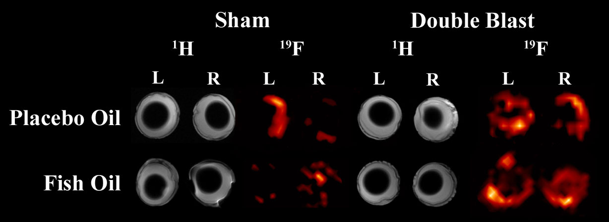
sham controls. Dietary omega-3 fatty acids (Fish Oil) have thus far shown
little, if any, ability to alleviate these acute injury events. Figure 1 shows
the increase in 19F signal in the eyes of rats exposed to double
blast compared to the sham rats and that treatment appears to have little or no
effect.
Discussion
These findings demonstrate that 19F MRI is effective in detecting systemic injury as a result of shock tube-generated overpressure. The injury itself is very heterogeneous, but one obvious result was that experiencing a second blast one minute after the first increased the amount of fluorine signal seen in these animals, which most likely represents increased inflammatory response (labelled macrophage accumulation), when compared with sham animals. Despite having potent anti-inflammatory properties, a diet rich in omega-3 fatty acids did not readily alleviate these acute injury events. Chronic events may be more amenable to other functions of omega-3 fatty acids, such as membrane structural restoration. More work needs to be done to explore the role that systemic inflammation has on not only acute and chronic injury, thus, we plan to extend evaluations over more prolonged post-blast intervals.
Acknowledgements
DoD grants from the MOMRP and USAMRMC/CDMRP, #: W81XWH-14-2-0178.References
1. Hanley, C. J. The Washington Post, http://www.washingtonpost.com. Accessed August 20, 2013.
2. Long JB, Bently TL, Wessner KA, et al. Blast overpressure in rats: recreating a battlefield injury in the laboratory. J Neurotrauma 2009; 26:827-840.
Figures