3588
A novel theranostic termoresponsive 19F probe for tumor imagingDaniel Jirak1, Andrea Galisova1, Ondrej Sedlacek2, Martin Hruby2, and Milan Hajek1
1IKEM, Prague, Czech Republic, 2Institute of Macromolecular Chemistry of the Academy of Sciences, Prague, Czech Republic
Synopsis
Novel 19F MR probe with the ability of creation of a depot in the living system is presented. This agent is pH- and termoresponsible with the phase transition point from liquid into the solid state at the temperature above 20 ºC. The MR properties were assessed by relaxometry and 19F spectroscopy/imaging. In vivo application confirmed efficiency of depot visualization; strong 19F MR signal was detected in all animals. No adverse effects of the probe to the animals were observed. Sufficient MR sensitivity of the probe and its slow degradation in animals suggest potential for a theranostic use.
Purpose
Fluorine 19F MR represents a promising tool for diagnostic imaging due to its high specificity caused by negligible abundance of fluorine in the tissues. Here we present a novel pH and temperature responsive 19F MR contrast agent based on fluorinated polymer systems. This agent allows phase transition from liquid into solid (gel-like) state at the temperature above 20 ºC. This forms an agent depot after injection into the living system, what could be very potential for tumor treatment after the drug incorporation. In this study, we have characterized the probe sensitivity and relaxivity dependence on temperature in the phantoms. Moreover, we have showed the depot in animal models in vivo for a long term.Methods
MR properties and responsivity to temperature of the fluorinated polymer systems (poly(2,2-difluoroethylacrylamide-co-1-imidazolylpropylacrylamide)) were tested. The temperature dependent MR properties of the probe were assessed by 1H and 19F relaxometry, 19F MR spectroscopy and 19F MR imaging. 1H relaxometry of the probe was performed on a 0.5T NMR relaxometer (r1 - saturation recovery sequence, recycle delay 12s; r2 – CMPG sequence, recycle delay 10s). Temperature-dependent 19F MR spectroscopy and imaging were carried out on a 4.7T scanner (MRS: single pulse, repetition time TR: 1000ms, number of acquisitions NA: 64, acquisition time TA: 1min; MRI: RARE sequence, TR: 2000ms, echo time TE: 7ms, turbo factor TF: 16, NA: 128, TA: 17min, resolution: 0.64x0.64x3 mm3). The sensitivity of the probe was assessed by 19F MR imaging at 25°C on a phantom containing various concentration of the probe (4.5 mM – 1.3 M). To check the feasibility of the 19F MR probe in vivo, four healthy female rats (Lewis, 250-300g) were scanned after the contrast agent administration (1.3 M, 300ml injection into the muscle of the right hind leg and into the subcutaneous area (left hind leg) for two months. Then we have administrated the agent (1.8 M, 200 mL) to the subcutaneous solid tumor (HUH7 cell line) and to the muscle of a CD-1 nude mouse. In animal experiments the same MR sequence as in the phantom study was used.Results
The relaxometry showed a strong relaxivity dependence on temperature, both relaxivity r1 and r2 were lower with increasing temperature (Fig. 1). Temperature-varied MRS and MRI experiment confirmed that dependence; we observed markedly lower MR signal in the phantoms with increasing temperature (Fig. 2). The minimal concentration of the agent at 25°C detected within reasonable measurement time for in vivo experiments (17 min) was 40 mM (Fig. 3). Strong 19F MR signal was detected at both injection sites in all animals; the 19F MR signal was detected two months after contrast agent administration and it was comparable with the signal detected in the first week (Fig.4). Signal to noise ratio (SNR) and volume of 19F MR signal after depot formulations within the first day slowly increased (Fig.5). No adverse effects of the probe to the animals were observed (no change of the body weight).Discussion
Good sensitivity and temperature-dependent nature of the novel 19F probe was confirmed. In the in vivo measurement, 19F MR signal enhancement after two hours following contrast agent administration reflects creation of a depot upon temperature change by overreaching the lower critical solution temperature. At temperatures above 20°C and pH = 7.4 contrast could change liquid phase to solid reversibly. Slow degradation confirmed also the result from volumetry; volume enlargement of depots between day 4 and day 57 after contrast agent application is probably due to slow diffusion of polymer depot to surrounding tissue. This feature, and the size of the nanoparticles (less than 200 nm) allowing spontaneous accumulation in solid tumors (enhanced permeation and retention (EPR) effect) might be attractive for the use as a theranostic tumor probe after drug incorporation.Conclusion
Here, we presented a novel efficient 19F MR probe which forms depot as a response to physiological parameters (pH and temperature) and can be detected in animals in vivo for a long term. Sufficient MR sensitivity of the probe and its slow degradation in animals suggest potential for theranostic use of this contrast agent.Acknowledgements
Supported by Czech Science Foundation GACR (P205-16-03156S), MH CZ-DRO (Institute for Clinical and Experimental Medicine–IKEM, IN 00023001).References
No reference found.Figures
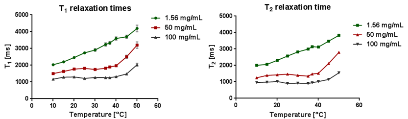
1H relaxation times dependence on temperature (1.29M, 0.645 M a 0.020 M
fluorine
concentration).
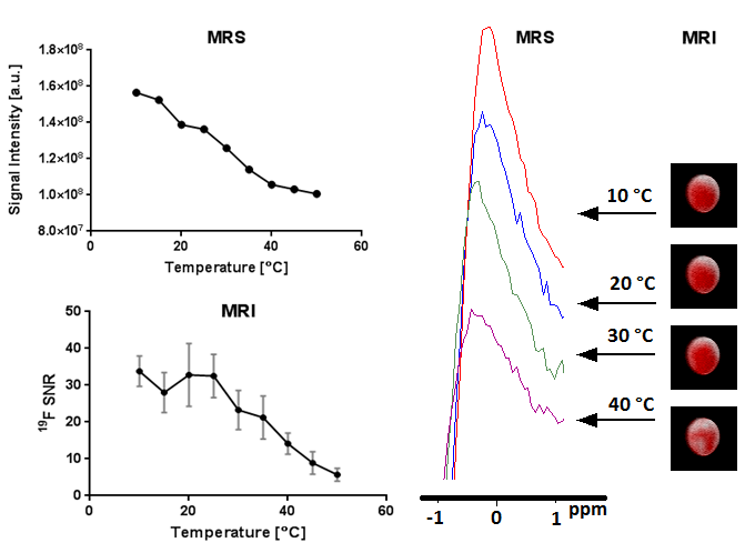
Dependence
of 19F MRS/MRI signal on temperature.
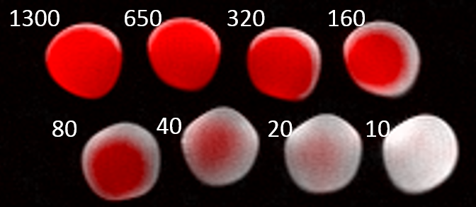
19F MR image of a phantom
overlaid on 1H MR image of the agent with different concentration;
the numbers represent concentration in mM.
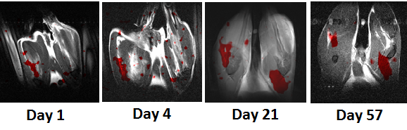
19F MR image (red color)
overlaid on 1H MR images of a rat with the injected agent into the
muscle (left) and subcutaneously (right). Days indicate day after the contrast
agent application.
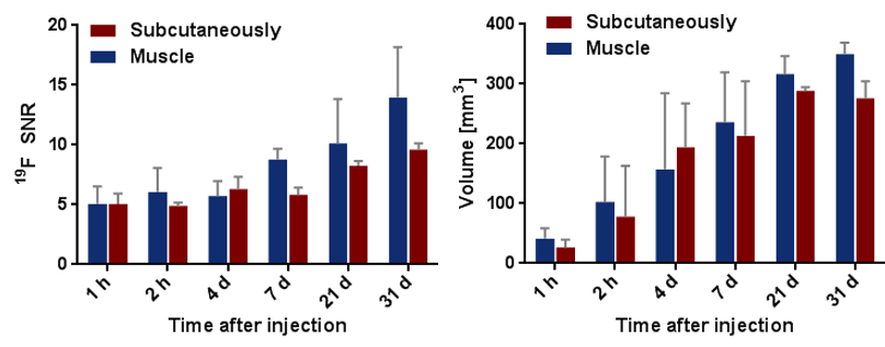
Signal
to noise ratio (SNR) (left) and volumetry (right) of polymer depots change during
the time after in vivo administration.