3584
Use of Gadofosveset-enhanced Lung MRI to Assess Ongoing Lung Injury in Fibrotic Interstitial Lung Disease.1Medicine, Pulmonary and Critical Care, Massachusetts General Hospital, Boston, MA, United States, 2Radiology, Massachusetts General Hospital, Boston, MA, United States, 3Siemens Healthcare, Boston, MA
Synopsis
Vascular leak is a cardinal response to injury, and prior assessments of alveolar-capillary permeability suggest that vascular leak is present in the lungs of patients, and that its extent is associated with progression and mortality. We hypothesized that the degree of vascular leak present reflects the extent of ongoing lung injury, and that measuring lung vascular permeability consequently could provide a much-needed metric for assessing ILD disease activity and predicting disease progression. Using magnetic resonance imaging with the albumin-binding contrast agent gadofosveset, we were able to detect increased vascular permeability in the lung of patients with fibrotic ILD.
Background
Fibrotic interstitial lung diseases (ILD) are often progressive with a high morbidity and mortality. Despite their overall poor prognosis, there is marked heterogeneity with some patients progressing rapidly while others have prolonged periods of clinical stability. At the current time neither assessment by computerized-tomography nor pulmonary function provide information as to how active an individual’s disease is at any one point in time – an urgent unmet clinical need. Vascular leak is a cardinal response to injury, and prior assessments of alveolar-capillary permeability suggest that vascular leak is present in the lungs of patients, and that its extent is associated with progression and mortality.1 We hypothesize that the degree of vascular leak present reflects the extent of ongoing lung injury, and that measuring lung vascular permeability consequently could provide a much-needed metric for assessing ILD disease activity and predicting disease progression at the time of diagnosis. Here we evaluated a novel method for evaluating lung vascular permeability using magnetic resonance imaging with the albumin-binding contrast agent gadofosveset. With this agent, tissue signal intensity is increased by prior extravasation of circulating endogenous albumin into tissues with vascular leak.Methods
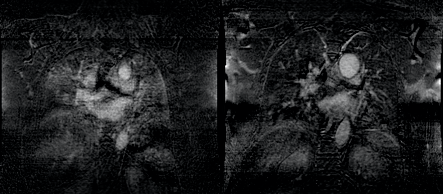
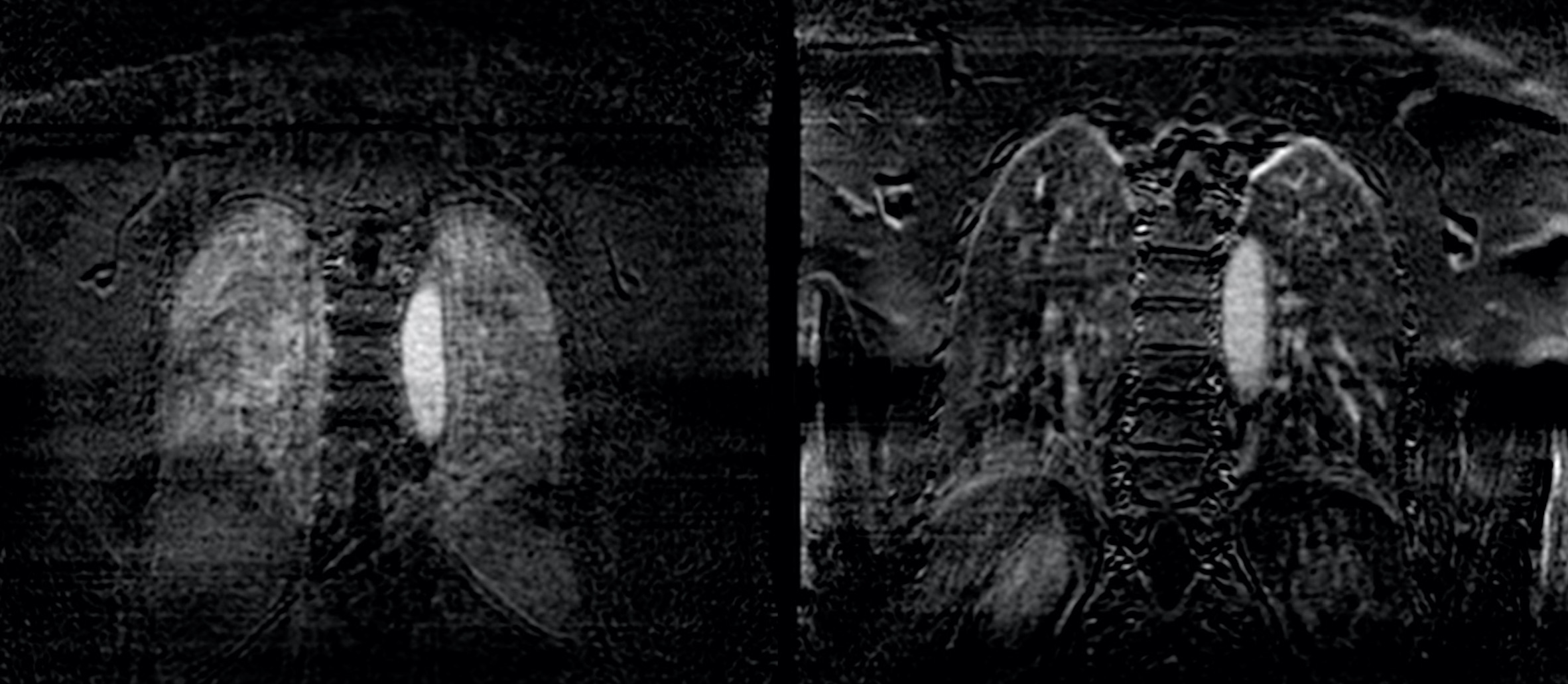
We performed gadofosveset-enhanced lung MRI on 5 healthy controls without known lung disease, and 7 patients with ILD, including 4 patients with idiopathic pulmonary fibrosis, 2 with non-specific interstitial pneumonia, and 1 with connective tissue-associated ILD. Participants were excluded for tobacco use within the prior 6 months, respiratory infection within the prior 6 weeks, congestive heart failure, renal impairment, or contraindications to MRI. MRI of the thorax was performed on a commercial 3T MRI scanner (PRISMA, Siemens Medical) using pointwise encoding time reduction with radial acquisition (PETRA). Images were obtained pre-contrast administration and at 2.5 to 5 minute intervals thereafter, up to 32.5 minutes after injection. Signal intensity was measured in standardized Regions of Interest (ROIs) in three regions (anterior, medial, and posterior) in the upper, middle, and lower lung parenchyma (see Figure 1), as well as three large vessels as control regions (ascending aorta, main pulmonary artery, and descending aorta), at each interval. Contrast reached steady state distribution 10 to 20 minutes post-injection. For each set of images acquired during this steady state, we calculated change in signal intensity in each parenchymal ROI (post minus pre-contrast level), divided by change in signal intensity in the control vessel region present in the same coronal image, i.e. the ascending aorta, main pulmonary artery, or descending aorta. Statistical analysis was performed using a student's t-test with p values </= being statistically significant.Results
Compared to the healthy controls, ILD patients had a significantly higher changes in lung MRI signal intensity following gadofosveset administration in multiple ROIs, including upper (left and right medial and posterior), middle (left anterior, right medial, left and right posterior), and lower (left anterior, right medial, and left and right posterior). Qualitative comparisons demonstrated increased vascular permeability with fibrotic ILD throughout all lung regions (Figures 1 and 2).Conclusions
Lung MRI represents a novel modality to image pulmonary vascular leak. Gadofosveset-enhanced lung MRI can quantitatively measure increased pulmonary vascular permeability in patients with ILD. As a marker of ongoing lung injury, measurements of vascular leak may be able to assess disease activity in patients with ILD, and predict their disease progression at the time of diagnosis.Acknowledgements
NIH/NHLBI 1F32HL129789-01 (Montesi, PI)
Harvard Catalyst Early Clinical Data Support for Grant Submissions (Montesi, PI)
References
1. Goh NSL, Desai SR, Anagnostopoulos C, et al. Increased epithelial permeability in pulmonary fibrosis in relation to disease progression. Eur Respir J 2011;38(1):184–90.Figures