3582
A high-relaxivity extradomain-B fibronectin targeting gadolinium metallofullerene for cancer detection and differential diagnosis in molecular MRIZheng Han1 and Zheng-Rong Lu1
1Case Western Reserve University, Cleveland, OH, United States
Synopsis
Accurate cancer detection and characterization with MRI is hampered by the lack of safe and effective targeted MRI contrast agents. In this work, we have designed and synthesized a high-relaxivity targeted contrast agent by conjugating ZD2 peptide to hydroxylated Gd3N@C80. ZD2-Gd3N@C80 has a high T1 relaxivity of 76.4 mM-1s-1 per Gd at 1.5 Tesla and a small diameter of 1 nm on average. At a low dose of 5 µmol Gd/kg, ZD2-Gd3N@C80 is able to produce prominent contrast enhancement in the highly metastatic MDA-MB-231 triple-negative breast cancer model, but not in the estrogen-dependent MCF-7 tumors.
Introduction
Accurate cancer detection and characterization of tumor aggressiveness has the promise to provide precision care to cancer patients. However, molecular MRI of cancer is hampered by the lack of safe and effective targeted MRI contrast agents with the ability of accurate cancer detection and differentiation of high-risk tumors from low-risk diseases. Currently available GBCAs suffer from low relaxivity and potential toxic side effects associated with the release of free Gd(III) ions. Recently, gadofullerenes have emerged as a novel class of MRI contrast agents with superior relaxivities1-3. In this work, we have developed a high-relaxivity targeted contrast agent by conjugating a small peptide ZD2 (Thr-Val-Arg-Thr-Ser-Ala-Asp) to hydroxylated Gd3N@C80. ZD2 specifically binds to EDB-FN highly expressed in many types of aggressive human cancer4. The targeted gadofullerene-based high-relaxivity contrast agent has the potential to address the low relaxivity and toxic side effects of the existing GBCAs and to enable accurate detection and risk-stratification of cancer with MRI at a much lower dose.Method
ZD2-Gd3N@C80 was synthesized by oxidation of Gd3N@C80 and subsequent peptide conjugation . To examine the relaxivity of the contrast agent, lyophilized ZD2-Gd3N@C80 was reconstituted to aqueous solutions in a concentration range of 0.0625 to 0.5 μM. T1 and T2 values of each solution were measured with a relaxometer (Bruker) at 1.5 T. The r1 and r2 relaxivities was calculated as the slops of the plots of 1/T1 and 1/T2 relaxation rates against the concentrations. Transmission electron microscopy (TEM) and dynamic light scattering (DLS) were used to characterize the morphology and size of the agent. The EDB-FN expression in MDA-MB-231 TNBC and estrogen receptor (ER) positive MCF-7 cells was determined using real-time PCR (RT-PCR) at the mRNA level. Mice bearing MDA-MB-231 and MCF-7 xenografts were used as high-risk and low-risk breast cancer models, respectively. An Aspect M3 MRI scanner (1 Tesla) was used for acquiring axial images of the mice at tumor locations. The axial T1 weighted tumor images were acquired with the following parameters: flip angle = 90°; TR = 500 ms; TE = 9 ms; field of view = 3 cm × 3 cm; matrix size = 128 × 128; slice thickness = 2 mm; inter-slice distance = 1 mm. Images were acquired at pre-injection, 10 min, 20 min, and 30 min after contrast injection. Tumor contrast-to-noise ratio (CNR) was calculated as tumor signal intensity subtracted by muscle signal intensity, scaled to noise.Results
The chemical structure of ZD2-Gd3N@C80 is shown in Fig. 1a. ZD2-Gd3N@C80 had high r1 and r2 relaxivities of 74.6 and 114.9 mM-1s-1 per Gd(III) ion, or 223.8 and 344.7 mM-1s-1 per molecule, respectively (Fig. 1b). Such superior relaxivities are critical to increase the sensitivity of contrast enhanced MRI for molecular imaging at low contrast agent doses. TEM and DLS indicated that ZD2-Gd3N@C80 had a small diameter of 1 nm on average (Fig. 1c and 1d), which is much smaller than the renal filtration threshold. As shown in Fig. 2a, cellular EDB-FN mRNA level in MDA-MB-231 cells was significantly higher than that in MCF-7 cells. MR images was acquired with the mice bearing MDA-MB-231 and MCF-7 tumor models before and after intravenous injection of ZD2-Gd3N@C80 at a very low dose of 1.67 µmol/kg or 5 µmol-Gd/kg, which is 20 times less than the dose of the clinical GBCAs. Quantitative analysis revealed that ZD2-Gd3N@C80 produced 39 to 45% increase of CNR in MDA-MB-231 tumors (n=4). No significant increase in tumor CNR was seen in MCF-7 tumors (n=3) (Fig. 2b and 2c).Discussion and Conclusion
Despite the efforts to increase the water-solubility of gadofullerenes to potentiate them for in vivo use1-3, little has been done in endowing tumor-homing abilities to these gadofullerenes. Here, we report the functionalization of a gadofullerene with an EDB-FN targeting peptide. The targeted contrast agent, ZD2-Gd3N@C80, has a high r1 relaxivity, which enables its use at a much lower dose to generate sufficient tumor contrast enhancement in MRI. The smaller size of ZD2-Gd3N@C80 also ensures fast clearance and fast tumor accumulation. Gd(III) ions are encapsulated in the fullerene cage, which prevents the release of toxic Gd(III) ions in the body. ZD2-Gd3N@C80 has the potential to provide accurate early detection and characterization of high-risk tumors and to minimize false-positive diagnosis with current MRI technology5. By stratifying the risk of cancer aggressiveness, molecular MRI with this contrast agent can assist the physicians to tailor personalized therapies for precision care of cancer patients.Acknowledgements
Research reported in this abstract was supported by the National Institute of Biomedical Imaging and Bioengineering of the National Institutes of Health under Award Number R01EB000489. The content is solely the responsibility of the authors and does not necessarily represent the official view of the National Institute of Health.References
1. Feng, Y. Q., Li, J., Zhang, Z. X., et al. A highly soluble gadofullerene salt and its magnetic properties. Dalton T 2015, 44, (17), 7781-7784. 2. Zhang, J., Ye, Y., Chen, Y., Pregot, C., et al. Gd3N@C84(OH)x: a new egg-shaped metallofullerene magnetic resonance imaging contrast agent. Journal of the American Chemical Society 2014, 136, (6), 2630-6. 3. Shu, C. Y., Zhang, E. Y., Xiang, J. F., et al. Aggregation studies of the water-soluble gadofullerene magnetic resonance imaging contrast agent: [Gd@C82O6(OH)(16)(NHCH2CH2COOH)(8)](x). J Phys Chem B 2006, 110, (31), 15597-15601. 4. Han, Z., Zhou, Z., Shi, X., et al. EDB fibronectin specific peptide for prostate cancer targeting. Bioconjugate Chem 2015, 26, (5), 830-838. 5. Baltzer, P. A., Benndorf, M., Dietzel, M., et al. False-positive findings at contrast-enhanced breast MRI: a BI-RADS descriptor study. AJR. American journal of roentgenology 2010, 194, (6), 1658-63.Figures
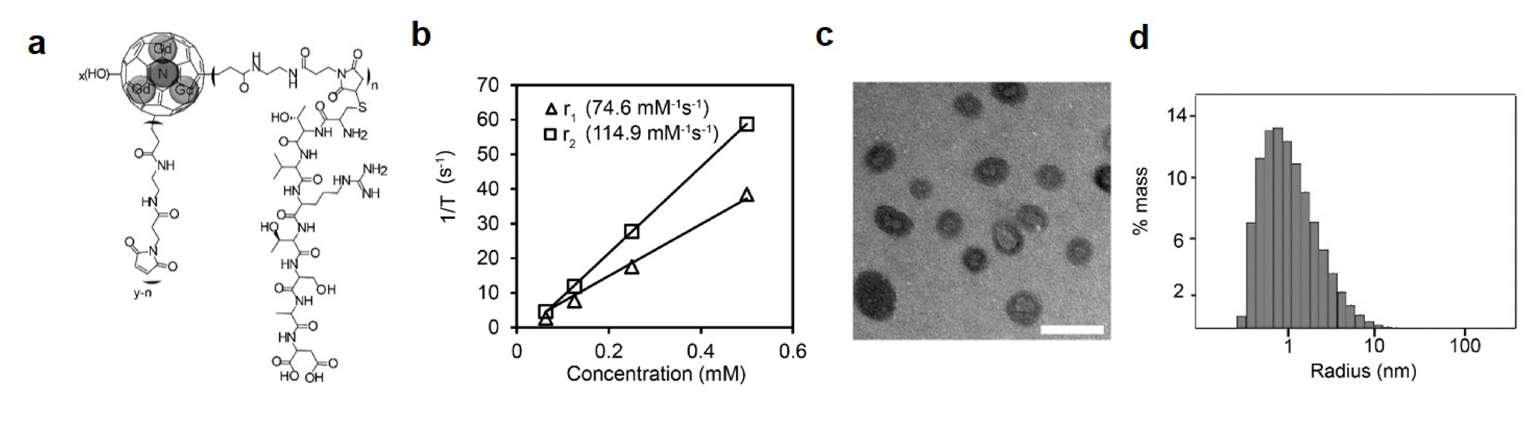
Fig.
1. a. Chemical structure of ZD2-Gd3N@C80. b. Plot of 1/T1 and
1/T2 versus ZD2-Gd3N@C80concentrations for calculation of r1
and r2 relaxivities. c. TEM images of ZD2-Gd3N@C80.
Scale bar: 5 nm. d. DLS analysis of ZD2-Gd3N@C80.
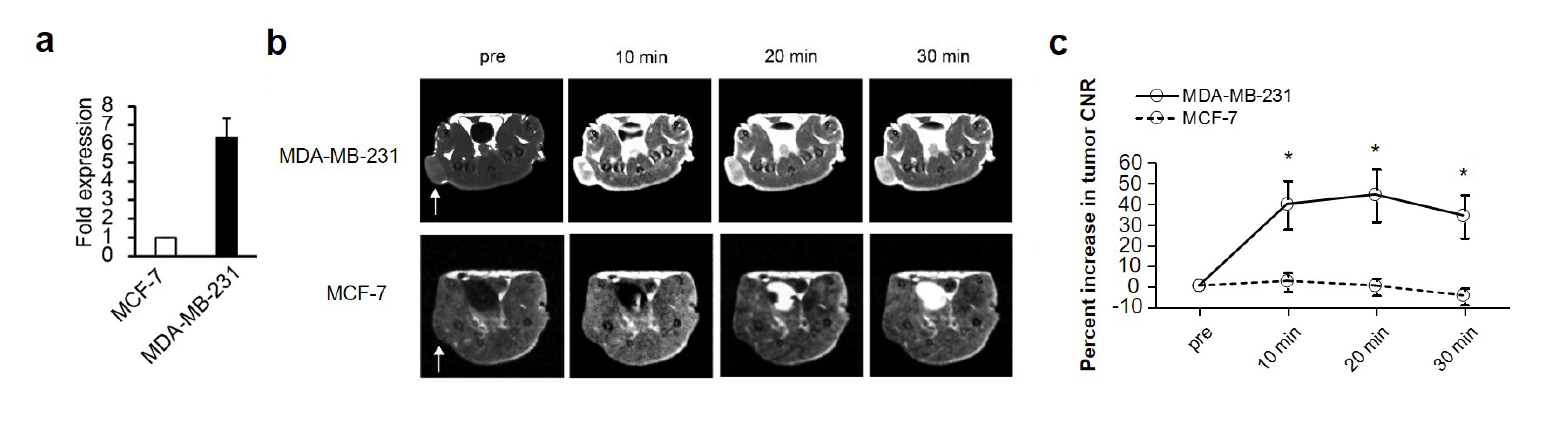
Fig.
2. a. RT-PCR analysis of EDB-FN
mRNA levels in MCF-7 and MDA-MB-231 cells, showing the higher EDB-FN expression
in MDA-MB-231 cells (n=3; P<0.05). The level of EDB-FN mRNA in MCF-7 cells
were used to normalize the data. b. Representative axial MRI images of
MDA-MB-231 and MCF-7 tumors. Images were acquired at 10, 20 and 30 min after
injection of ZD2-Gd3N@C80, Tumor locations were indicated
by white arrows. c. Quantification of tumor CNR after contrast agent injection.