3579
PRO-QUEST: A fast method for exchange rate quantification based on PROgressive saturation for Quantifying Exchange using Saturation Times in Chemical Exchange Saturation Transfer (CEST)1Brain Repair and Rehabilitation, Institute of Neurology, London, United Kingdom, 2Department of Medical Physics & Bioengineering, University College of London, London, United Kingdom
Synopsis
In this work we propose a novel pulse sequence for measuring chemical exchange rates through a progressive saturation recovery process PRO-QUEST (PROgressive saturation for Quantifying Exchange using Saturation Times). The water magnetization is sampled under non-steady state conditions and saturation is interleaved with the acquisition of images obtained in a single scan. Unlike previous approaches, it provides maps of T1 and B1 , needed for exchange-rate calculations, from the same dataset. The proposed pulse sequence has been successfully applied to obtain exchange rate maps in phantoms and healthy rat brains.
Introduction
While different methods have been proposed to enable correct exchange rate estimates, they require so much time that their utility has not really been explored in the clinic1,2. In this work we aimed to develop a method to make rapid and accurate measurements of exchange rates through a progressive saturation recovery process. To achieve this, we modified a Look-Locker sequence, used primarily for T1 mapping3, in such a way that an off-resonance saturation pulse or a spin-lock module was interleaved with the acquisition of exchange weighted images. We sampled the signal evolution with and without saturation pulses and derived mathematical equations to enable calculations of T1, B1 and exchange rates from the same dataset with an acquisition time of only 16 minutes.Methods
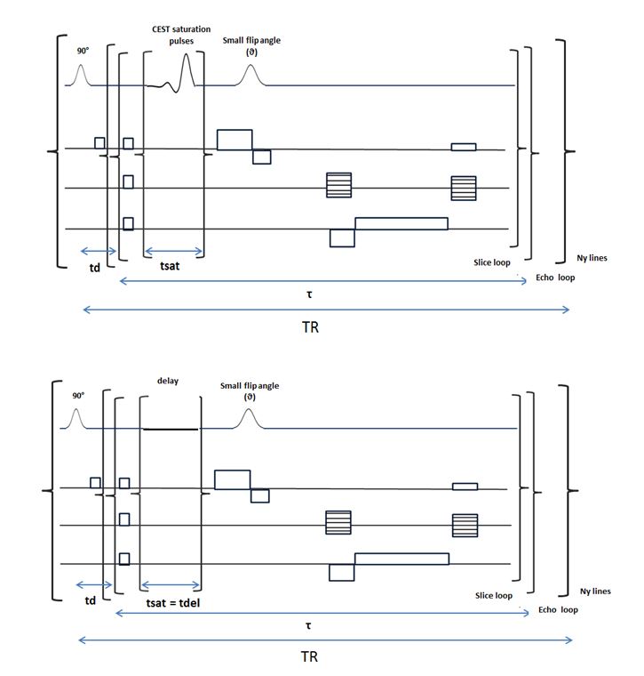
The PRO-QUEST sequence was implemented on a 9.4 Tesla Agilent MRI scanner. A detailed diagram of the pulse sequence is displayed in figure 1. Each of the n=300 off-resonance saturation pulses was centered at 3.0 ppm with parameters: pulse duration = 6.23 msec, flip angle = 90° and bandwidth = 1000 Hz. Three additional scans were acquired, with the saturation pulses being replaced with delays, at three different imaging flip angles =8°, 15° and 25 ° for estimating T1, B1 and Mo similarly to previous work4.For readout, a single-slice 2D-GRE sequence with FOV 20x20 mm data matrix 64x64, TR=5 sec and TE=1.3 msec was employed. The slice thickness was 4 mm. PRO-QUEST measurements were made in phantoms containing 100mM of Alanine at pH=5.9, 6.2, 6.4 and 7.0 using a transmit/receive RF coil with 33mm inner diameter (all RF coils: Rapid Biomedical, Germany).
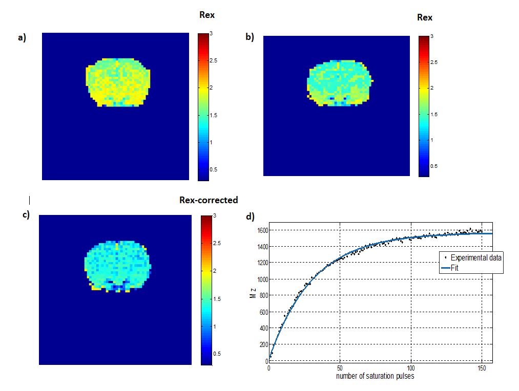
To demonstrate the feasibility of obtaining exchange rate measurements in vivo using the PRO-QUEST sequence, 3 healthy DA rats were scanned using a volume coil with an inner diameter of 72 mm for radio frequency (RF) transmission and a two-element receive array coil. The optimized sequence consisted of two adiabatic half-passage saturation pulses for achieving water suppression followed by 150 off-resonance saturation pulses centered at 2.0 ppm with pulse parameters: pulse duration 16.93 msec, flip angle = 90° or 180° and bandwidth = 400 Hz. Images were collected with a slice thickness = 2 mm, FOV = 35 x 35 mm, matrix size=64x64, TR = 3.9 sec, TE = 1.12 msec, and imaging flip angle 25°. To improve the image SNR two averages were used. In addition, T1, B1 and M0 were calculated using the multiple flip angle strategy described above (i.e. 8°,15° and 25 °).
Data processing was performed using custom-written scripts in MATLAB (Mathworks, Waltham, MA). The progressive saturation recovery curves as a function of delays (defined in figure 2) were fitted to the experimental data to provide measurements of T1 (=1/R1), B1 (related to θ)4 and M0 using the following equation3:
Mzd (nτ)= (1-[(cosθ)n exp(-nτR1) ])/(1-[(cosθ)exp(-τR1)]) Mzd (τ) + M0 (1-exp(-td R1))[ (cosθ)n ) exp(-n(τR1 ) )] [1]
The obtained T1, ϑ and M0 values were then used as input parameters for estimating the exchange rate by fitting the experimental data to our derived equation:
Mzsat (nτ)= (1-[α(cosθ)n ) exp(-n(τR1-tsat (R1-R1ρ )))])/(1-[α(cosθ) exp(-(τR1-tsat (R1-R1ρ )))]) Mzsat (τ)
+ M0 (1-exp(-(tdR1)))[ (cosθ)n) exp(-n(τR1-tsat (R1-R1ρ)))] [2]
where Mzsat (τ) = Mss (1- exp(-(R1ρ tsat ))) cosθ exp(-((τ-tsat)R1 )) + M0 (1-exp(-(τ-tsat)R1 )) and R1ρ=R1+Rex
Results
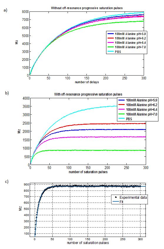
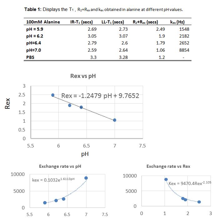
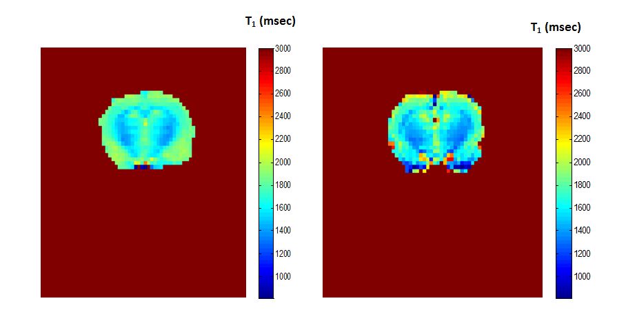
Progressive saturation recovery curves with and without saturation pulses in Alanine at various pH values are shown in Figure 2. The calculated Rex5 and exchange rates for all the samples are displayed in table 1 with calculated calibration curves in Figure 3. In addition, the T1 values using both the saturation recovery LL sequence (SR-LL) and an Inversion recovery EPI sequence (IR-EPI) are displayed for comparison. Figure 4 shows the T1 maps calculated in a healthy rat brain using both IR-EPI and SR-LL sequences. The Rex maps at off-resonance pulse power levels of 2.4 μT and 4.8 μT and their ratio which it is independent of concentration are shown in Figure 5.Discussion and conclusion
We have implemented a new pulse sequence for exchange rate quantification and pH mapping called PRO-QUEST. Validation experiments were performed in phantoms with the exchange rates of amine protons found to increase with pH, in line with the literature6. Furthermore, the new sequence was successfully applied to estimate Rex in the healthy rat brain. In future, we aim to apply PRO-QUEST for pH mapping and exchange rate quantification in stroke or tumours.Acknowledgements
No acknowledgement found.References
1. McMahon M,Assaf A. Gilad,Zhou J et al. Quantifying Exchange Rates in Chemical Exchange Saturation Transfer Agents Using the Saturation Time and Saturation Power Dependencies of the Magnetization Transfer Effect on the Magnetic Resonance Imaging Signal (QUEST and QUESP): pH Calibration for Poly-L-Lysine and a Starburst Dendrimer. Magn Reson Med. 2006 April ; 55(4): 836–847.
2. Dixon WT, Ren JM, Lubag AJM, et al., A Concentration-Independent Method to Measure Exchange Rates in PARACEST Agents. Magnetic Resonance in Medicine. 2010; 63(3):625–632.
3. D.C. Look and D.R. Locker, “Time Saving in Measurement of NMR and EPR Relaxation Times”, Review of Scientific Instruments 41, 250 - 251 (1970).
4. D. L. Parker, B.A. Christian, K.C. Goodrich, et al. Improved accuracy in T1 measurements. In Proceedings of the 6th Annual Meeting of ISMRM,Sydney, Australia 1998 p. 2172.
5. Jin T, Autio J, Obata T, et al. Spin-locking versus chemical exchange saturation transfer MRI for investigating chemical exchange process between water and labile metabolite protons. Magn Reson Med. 2011 May;65(5):1448-60.
6. Zong X., Wang P, Kim S, et al. Sensitivity and Source of Amine Proton EXchange (APEX) and Amide Proton Transfer (APT) MRI in Cerebral Ischemia. Magn Reson Med. 2014 January ; 71(1)
Figures