3562
Retrospective reconstruction of hepatic arterial phases in gadoxetic acid-enhanced liver MR using continuous golden-angle radial sequenceYoon-Chul Kim1, Eunju Kim2, Gabrielle Beck3, Hans Peeters3, and Young Kon Kim4
1Clinical Research Institute, Samsung Medical Center, Sungkyunkwan University School of Medicine, Seoul, Korea, Republic of, 2Philips Healthcare, Seoul, Korea, Republic of, 3Philips Healthcare, Best, Netherlands, 4Radiology, Samsung Medical Center, Seoul, Korea, Republic of
Synopsis
We apply a 3D continuous golden-angle radial sparse parallel (GRASP) sequence to gadoxetic acid-enhanced imaging of the liver at 3 T. Arterial phase imaging may need high spatial and temporal resolution as well as correct timing of capturing arterial enhancement to obtain hemodynamic information of the tumor. Continuous golden-angle radial sampling provides a flexible retrospective selection of temporal window. We develop a technique that identifies the arterial peak from high temporal resolution data and reconstructs arterial phases with different temporal windows. The characteristics of hepatic arterial images after retrospective temporal resolution selection are demonstrated.
Introduction
Conventional breath-hold single phase imaging in gadoxetic acid-enhanced liver MR is known to produce artifact in hepatic arterial phase images due to transient dyspnea. Triple arterial phase imaging can acquire an arterial phase image unaffected by severe motion, but data acquisition results in lower SNR due to reduced temporal window1. Free-breathing and continuous acquisition using golden-angle radial sparse parallel (GRASP) is an alternative and has the potential to reduce motion artifact in arterial phase imaging2. Accurate timing for capturing arterial phase is important in reconstruction of continuously acquired data. In this abstract we study a retrospective reconstruction technique that first identifies the timing of contrast enhancement, adjusts time offset in raw data, and reconstructs arterial phase images with different temporal windows.Methods
Gadoxetic acid-enhanced liver MRI data were acquired during free breathing using a 3D stack-of-stars golden-angle radial sequence on a Philips Achieva 3T scanner. Sequence parameters were as follows: scan time = 1:04, TR/TE = 3.5/1.51 ms, total radial spokes = 379, partial k-space along slice encode = 41/52, flip angle = 10°, image matrix = 504 x 504 x 52, reconstruction voxel size = 0.73 x 0.73 x 2.0 mm. We scanned five healthy volunteers, and the MRI protocol included other routine abdominal MR sequences: T2 weighted imaging, diffusion weighted imaging, eTHRIVE DCE imaging, etc. The BART parallel imaging compressed sensing reconstruction was adopted for the improvement of spatial and temporal resolution and was implemented in MATLAB3. (Identification of maximal aortic enhancement frame) We reconstructed a single slice with temporal resolution of 21 spokes (i.e., 3.5 sec) and measured average signal in the aorta region of interest (ROI) at every frame. The time frame corresponding to the maximum aorta signal served as a reference for determination of arterial phase images. (Retrospective reconstruction with different temporal windows) Based on the peak enhancement frame, we adjusted time offset in the raw data and reconstructed images using parallel imaging compressed sensing. Temporal windows of 55 and 70 contiguous spokes were considered for image quality comparison. Temporal total variation was used for reconstruction.Results and Discussion
The aorta ROI is shown in Fig 1a. The time intensity curve shows the pattern of contrast enhancement followed by contrast decay (Fig 1b). Although the 21-spokes reconstruction is highly under-sampled, it produces acceptable image quality in the aorta ROI and enables the identification of peak arterial enhancement with high temporal resolution. The peak enhancement frame served as a reference time point. Based on the reference time point (see the red dashed line in Fig 1c), we assigned the temporal frames in the raw data. Fig 2 shows comparison of different temporal resolution reconstructions from the same continuously acquired raw data. The 55-spokes reconstruction shows that the enhancement of hepatic vessel first occurs in 10.1 – 20.2 seconds while the 70-spokes reconstruction does exhibit the enhancement pattern in 0-12.8 seconds. This indicates that the enhancement in the hepatic vessel starts to occur between 10.1 and 12.8 seconds. The 70-spokes reconstruction suggests that higher SNR image can be obtained with longer temporal window in late hepatic arterial phase in which its contrast change is relatively slower than early arterial phase. In this work, we have not addressed artifact issue resulting from respiratory motion. We will incorporate a self-gating technique4 into reconstruction and study its effect on image quality.Conclusion
We have demonstrated that the identification of peak aortic enhancement is feasible with high temporal resolution reconstruction of continuously acquired golden angle radial sequence data. The peak enhancement time information can provide a reference time point. Subsequent retrospective reconstructions with different temporal windows offer flexibility in selecting images from early arterial and late arterial phases.Acknowledgements
No acknowledgement found.References
[1] Pietryga et al., Radiology 2014:271(2):426-434. [2] Chandarana et al., Investigative Radiology 2013:48(1):10-16. [3] Uecker et al., ISMRM 2015 p2486. [4] Feng et al., MRM 2016:75:775-788.Figures
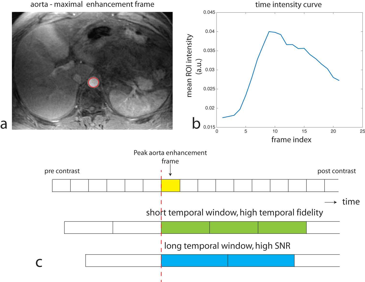
Fig 1. Retrospective
determination of peak aortic enhancement timing. (a) ROI of the aorta (red).
(b) Time intensity curve of the mean aorta signal. Maximum frame index is
identified as 9. (c) Illustration of retrospective reconstruction of arterial
phase images from continuously acquired golden angle data. Start time of the
raw data was aligned in reconstructions of different temporal windows.
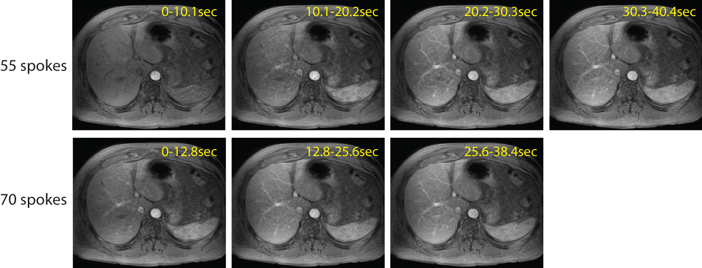
Fig 2. Retrospective reconstructions of
contiguous time frames in two different temporal windows. (Top) 55 spokes. (Bottom)
70 spokes. The 55-spokes result shows
temporal differentiation of early arterial enhancement. The 70-spokes result
shows better SNR in late arterial phase images (compare 20.2-30.3sec image and
25.6-38.4sec image).