3553
Evaluation of T1 Parametric Mapping using Inversion Recovery Fast Spoiled Gradient Echo: Application for Pre- and Post-Contrast Liver MRI1Department of Radiology and Imaging Sciences, Emory University, Atlanta, GA, United States, 2MR R&D Collaborations, Siemens Healthcare, Atlanta, GA, United States, 3MR Application Predevelopment, Siemens Healthcare, Erlangen, Germany
Synopsis
This investigation evaluates the performance and accuracy of a fast inversion recovery Look-Locker method for in vivo liver T1 mapping. Several parameters were assessed using T1 phantoms to describe accuracy trends and prevalence for artifacts. The method was also applied in vivo to demonstrate feasibility of fast T1 mapping of liver parenchyma and blood pool. The results offer insight into optimal imaging parameters, and showed good agreement with known T1 values at 1.5T.
Introduction
Recently, quantification of T1 using fast MRI methods has been a promising surrogate for assessing liver fibrosis both pre- and post-Gd contrast1,2. Even though ECG-gated sequences, such as the modified Look-Locker (MOLLI) method, have been adapted for body imaging, there has been limited evaluation of un-gated fast gradient-echo Look-Locker T1 mapping (LL-T1), which acquires continuous longitudinal recovery data following an inversion (IR) pulse3, especially for optimizing liver application. Therefore, the purpose of this study was to investigate the performance and accuracy of LL-T1 using MR phantoms, and to demonstrate feasibility of in vivo T1 mapping of liver parenchyma and blood pool.Methods
All MR imaging was performed at 1.5T (Magnetom Aera and AvantoFit, Siemens Healthcare, Erlangen, Germany). Ten MR phantoms were created, containing a homogeneous composition of either 1% (n=3) or 2% (n=7) agar gel and varying concentrations of gadobenate dimeglumine (Gd-BOPTA, Multihance, Bracco, Italy): 0, 0.5, 1.0mM (1% agar); 0, 0.1, 0.2, 0.3, 0.4, 0.5, 1.0mM (2% agar). Reference T1 measurements were measured using an IR spin-echo sequence (TR/TE=8000/6.3ms) with 13 TIs spanning 50 to 3400ms. Thereafter, a prototype LL-T1 gradient-echo sequence was applied with the following parameters: FOV=300x235mm; matrix=384x240 (interpolated); TR/TE/flip = 2.3/0.9ms/8; BW=1185Hz/px; GRAPPA=2. Three key parameters were systematically varied for optimization: number of TI images (#TIs=16, 24, 32, 48, 64), k-space segments (segs=1, 2, 3, 6), and the “free relaxation period” between IRs (TF=2, 3, 4, 6sec). Inline T1 maps were generated using prior fitting methods3, and mean T1 and standard deviation (SD) were recorded. Accuracy was determined by absolute % difference with reference T1, while performance was assessed using a coefficient of variation (CoV=SD/mean) and the presence of artifacts. For comparison, two MOLLI methods (using SSFP) were also acquired using timing schemes4: 5(3)3 and 4(3)3(3)2. Statistical comparisons were made using a two-tailed t-test, with p=0.05. For initial clinical feasibility, LL-T1 was performed in 11 prospective patients both pre- and post-contrast (>5min post Gd-BOPTA) using specific parameter variations (#TIs=32 or 48; segs=1 (n=10); #TIs=16, segs=2, TF=3s (n=1)) and similar imaging parameters as the phantom analysis. Mean T1 and SD was measured from 3 liver (n=33) and 1 aortic blood (n=11) locations, and compared to known values at 1.5T. Presence of artifacts was also observed.Results
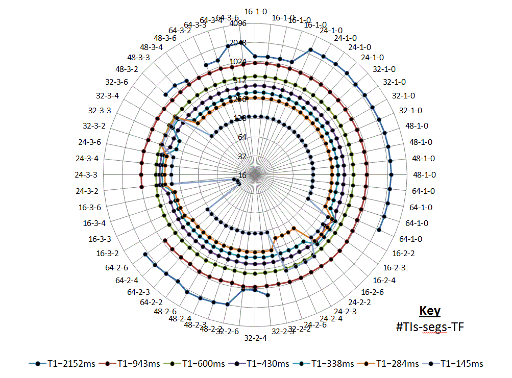
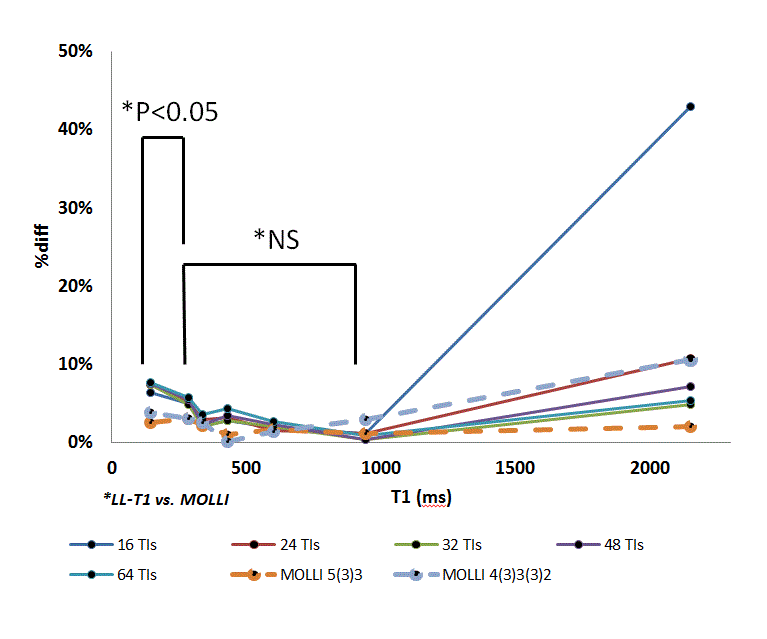
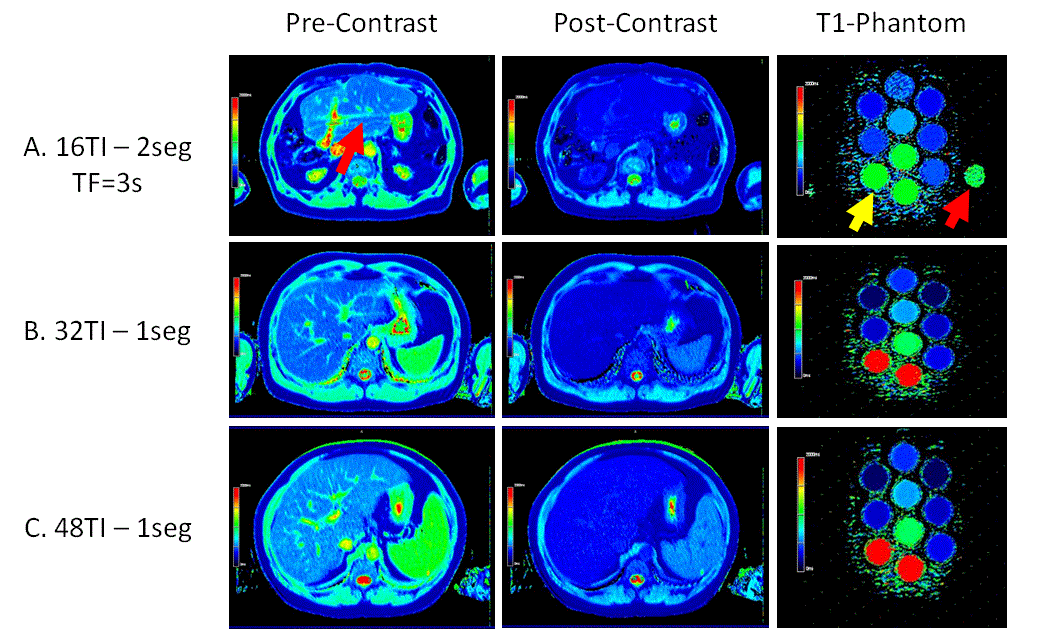
Due to failed inline T1 fitting, some data (segs=6) were omitted from analysis. Figure 1 depicts the T1 results and performance trends of all 2% agar phantom data using a log2 radar plot for improved visualization (1% agar showed similar results). Poor T1 fitting, signal fluctuations, and the presence of phase ghosts were observed on long T1 phantoms using LL-T1 with segs=3, or low TFs (<4s) and low #TIs (<24) using segs=2. In general, single-shot (segs=1) LL-T1 with #TIs>16 was associated with the lowest %difference (< 5% error) and CoV (< 0.04) over the T1 range. Single-shot LL-T1 is depicted in Figure 2, along with MOLLI results, showing equivalent performance between the methods for T1 spanning approximately 250 and 1000ms (p>0.05). However, CoV was lower for MOLLI (<0.01) than LL-T1 (>0.02) over this range. Improvement in T1 accuracy and CoV was not observed for increasing #TIs from 24 to 64, except for estimating long T1 (>1000ms). Figure 3 shows phantom and in vivo LL-T1 images, along with ghost artifact sensitivity for LL-T1 with segs=2 and TF=3s. Average T1 in both liver and blood was not different between #TIs=32 and 48 LL-T1 methods. Pooled pre-contrast T1 was 532.6 ± 37.9ms and 1380.7 ± 181.1ms for liver and blood, respectively, while post-contrast T1 was 331.5 ± 34.5ms and 388.7 ± 66.3ms, respectively. Average pre-contrast liver and blood T1 closely matched literature values5.Discussion
The improved performance of single-shot LL-T1 reflects the lack of a prescribed wait period (TF) between IR preparations, which may cause signal fluctuation in k-space. Though segmented LL-T1 can be performed with #TIs>32 and TF>4s, the associated scan time precludes acceptable in vivo breath hold duration. Single-shot LL-T1 with #TIs>24 performed best over a broad range of T1, despite some discrepancy with MOLLI for low T1 estimation. This relative inaccuracy may reflect the greater T1 signal variation during GRE data acquisition (LL-T1) compared to SSFP (MOLLI). Though in vivo T1 measures match literature values, blood T1 measures varied noticeably due to flow effects.Conclusions
This study provides a comprehensive assessment of IR Look-Locker imaging in phantoms, and initial application for liver T1 mapping. For broad T1 accuracy and low CoV, single-shot LL-T1 with #TIs>24 was optimal. Further in vivo validation is necessary against other accepted T1 methods.Acknowledgements
No acknowledgement found.References
1. Pavlides, M., Banerjee, R., Sellwood, J., et al. Multiparametric magnetic resonance imaging predicts clinical outcomes in patients with chronic liver disease. Journal of Hepatology. 2016;64, 308-315
2. Yoon, J. H., Lee, J. M., Paek, M., et al. Quantitative assessment of hepatic function: modified look-locker inversion recovery (MOLLI) sequence for T1 mapping on Gd-EOB-DTPA-enhanced liver MR imaging. European Radiology 2016;26, 1775-1782
3. Deichmann R, and Haase A. Quantification of T1 Values by SNAPSHOT-FLASH NMR Imaging. Journal of Magnetic Resonance. 1992; 96, 608-612
4. Kellman, P., and Hansen, MS. T1-mapping in the heart: accuracy and precision. Journal of Cardiovascular Magnetic Resonance 2014;16, 1-20.
5. Stanisz, GJ., Odrobina, EE., Pun, J., et al. T1, T2 relaxation and magnetization transfer in tissue at 3T. Magnetic Resonance in Medicine 2005;54, 507-512
Figures