3549
Reliable determination of bile acids from human gallbladder by 1H MRS - protocol optimization and estimation of reproducibility1Depts. Radiology and Clinical Research, University Bern, Bern, Switzerland, 2Dept. Visceral Surgery and Medicine, University Inselspital, Bern, Switzerland
Synopsis
Bile exerts multiple functions in the liver and gut with a crucial role for triglyceride-, sterol- and carbohydrate-metabolism and is a key player in disease processes. The study purpose was to develop a reliable MRS protocol and to assess variability of bile acid determination in human gallbladder. Our study demonstrated higher stability and reliability of gallbladder spectra with subjects measured in prone position compared to back position. Relatively small coefficients of variation were obtained in a reproducibility study particularly within subjects, suggesting clinical applicability of the method, especially for longitudinal studies.
Introduction
Bile exerts multiple functions in the liver and gut playing a crucial role for triglyceride-, sterol- and carbohydrate-metabolism. Moreover, bile acids influence nutrient metabolism and energy expenditure and have been evidenced to be potent modulators of the intestinal flora, integrity and inflammation. Therefore, bile is a key player in disease processes including liver regeneration, hepatic and colonic cancer, inflammatory bowel disease and metabolic syndrome. The bile in the gallbladder closely reflects the bile acid pool available and being secreted into the gut. Therefore, our long-term aim is to establish a diagnostic method utilizing 1H-MR spectroscopy (MRS) in determining bile composition of the gallbladder non-invasively. Only two studies have been performed previously investigating human bile from gallbladder in-vivo using 1H-MRS1,2, including an excellent study acquiring 1D- and 2D-spectra2. The lack of studies is presumably due to difficulties in obtaining reliable spectra in a small and moving organ, which additionally often suffer from susceptibility differences from bowel gas. To our knowledge, reproducibility, normal variation, and measurement success rate have not been determined yet. The purpose of this study was therefore to develop a reliable MRS protocol and to assess variability of bile acid determination in human gallbladder, which are preconditions for potential clinical studies.Subjects & Methods
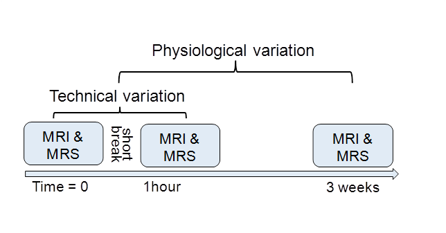
Study Population: Twenty-four measurements were performed on 17 healthy volunteers for protocol optimization. In addition, six healthy volunteers were measured three times: once back-to-back leaving the magnet in between measurements and once again after approximately 3 weeks to assess the technical and physiological variability, respectively (Fig.1). All measurements were performed after over-night fast on a 3T-MR Scanner (Verio,Siemens) with the subjects either in back or in prone position.
MR Spectroscopy: A single-voxel PRESS sequence with PACE-triggering (TR= 1 respiration cycle, TE=35ms, 16measurements with 4 acquisitions, 6 saturation bands, voxel size between 12x12x12mm3 and 15x15x15mm3 adapted to the individual gallbladder size) was acquired with the voxel placed in the center of the gallbladder. MRS measurement duration was about 5min, depending on breathing cycle. A non-water-suppressed spectrum was acquired for quantification2.
Data Processing: Spectra were summed after inspection and potential discard of individual spectra. Fitting of the bile acid and lipid peaks from the summed spectrum was performed using jMRUI AMARES3. The results were normalized to the non-water-suppressed spectrum.
Ex-Vivo bile measurements: For metabolite assignment and to obtain prior knowledge for the fitting of in-vivo spectra, ex-vivo human bile was measured on a 500MHz Bruker spectrometer, using a water-suppressed PROJECT4 sequence. Bruker-TopspinTM software was used for spectral processing.
Statistical analysis: Coefficients of variation between (CVB) and within (CVW) subjects were calculated for the metabolites. CVB and CVW were calculated for both, back-to-back scans (CVB_techn., CVW_techn.) and for scans separated by 3 weeks (CVB_physiol., CVW_physiol.).
Results & Discussion
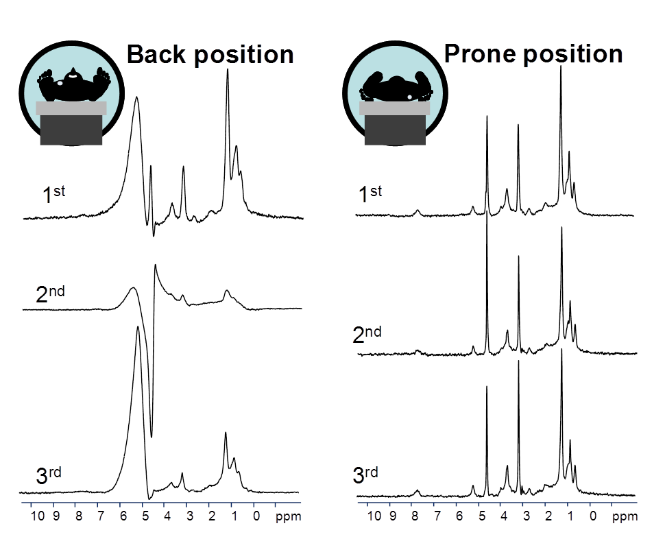
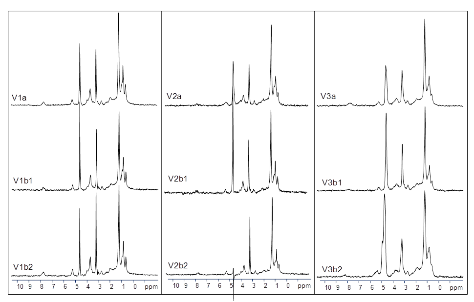
Protocol optimization: Gallbladder measurements with the subjects lying on their back resulted in a low measurement success rate of <50% acceptable spectra (based on linewidth, lipid contamination, or spurious signals, Fig.2-left), This is possibly mainly due to the superficial location of the gallbladder, which is susceptible to respiratory movements especially in the back position, as well as due to occasional susceptibility differences at tissue-air interfaces due to bowel gas. In contrast, measuring the volunteers in prone condition provided stable and reliable results: No subject measurement had to be excluded from further analysis (success rate 18/18 spectra, Fig.2-right). Average linewidth of the trimethylammonium-peak from choline-containing phospholipids at 3.23ppm was 8.0±3.7Hz (range:4.5-19.9Hz, Fig.3) for the 18 measurements in prone position. This high success rate is probably due to almost complete prevention of respiratory movement of the gallbladder in prone position.
Reproducibility: The 3-times repeated measurements on six subjects in prone position allowed to determine technical and physiological variations (Fig.1). Examples of repeated spectra of three volunteers (Fig.3) demonstrate visually good spectral quality and reproducibility.
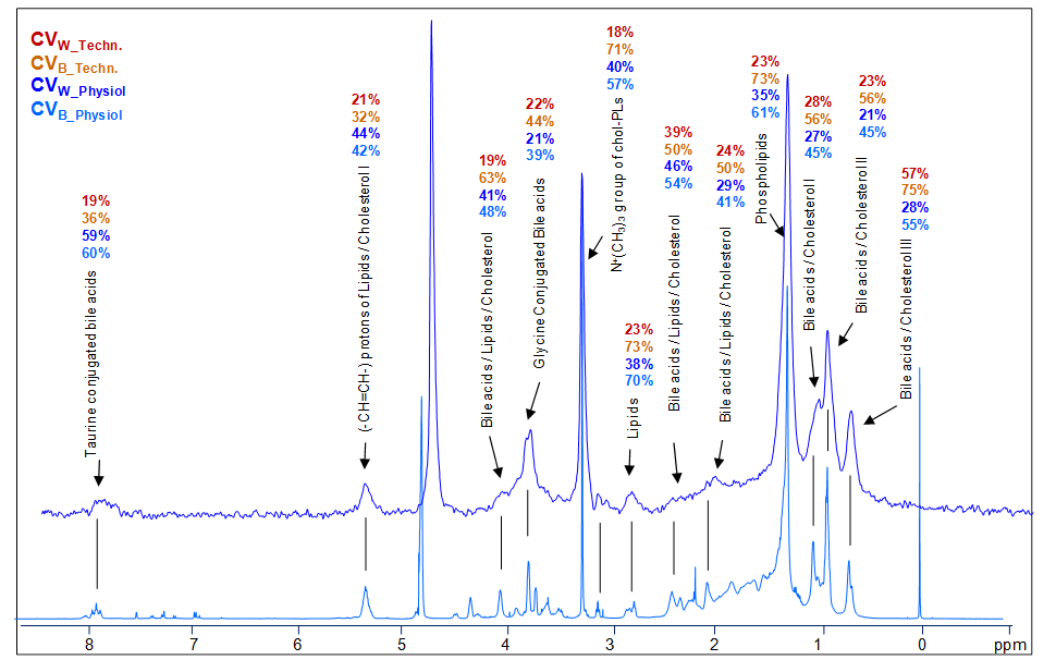
Peak assignments are indicated in Fig.4 on an in-vivo example spectrum from the gallbladder and an ex-vivo human bile spectrum, together with the determined CVW and CVB for the main determined metabolites. Relatively low CVW-values were obtained for main metabolites with most values between 20-30% for both, the back-to-back measurements and for the 3 weeks separated scans. In contrast much higher values were obtained for CVB. The variance was not significantly different between the back-to-back scans and the physiological replicates.
Conclusion
Our study demonstrated higher stability and reliability of gallbladder spectra with subjects measured in prone position compared to back position. Therefore, for gallbladder spectroscopy measurements, prone position of the subject is suggested. Moreover, the obtained relatively small coefficients of variation particularly within subjects suggest clinical applicability of the method, especially for longitudinal studies.Acknowledgements
This work was supported by the UniBE ID-Grant (PV).References
1. Prescot AP, Collins DJ, Leach MO, Dzik-Jurasz AS. Human gallbladder bile: noninvasive investigation in vivo with single-voxel 1H MR spectroscopy. Radiology 2003;229:587-592.
2. Mohajeri S, Ijare OB, Bezabeh T, King SB, Thomas MA, Minuk G, Lipschitz J, Kirkpatrick I, Smith M, Smith IC. In vivo 1H MRS of human gallbladder bile at 3 T in one and two dimensions: detection and quantification of major biliary lipids. NMR Biomed 2014;27:1192-1202.
3. Vanhamme L, van den Boogaart A, van Huffel S. Improved method for accurate and efficient quantification of MRS data with use of prior knowledge. J Magn Reson 1997;129:35-43.
4. Aguilar JA, Nilsson M, Bodenhausen G, Morris GA. Spin echo NMR spectra without J modulation. Chemical Communications 2012;48:811-813.
Figures