3544
Feasibility of computed diffusion-weighted MR imaging for visualization of pancreatic adenocarcinoma: comparison with acquired diffusion-weighted imaging1Radiology, Kagoshima University Graduate School of Medical and Dental Sciences, Kagoshima, Japan, 2Philips Electronics Japan, TN, Japan
Synopsis
We compared directly acquired diffusion-weighted imaging (DWI) at b-values of 1500 s/mm2 and 2000 s/mm2 with computed DWI (cDWI) at the same b-values, which was calculated from directly acquired DWI data at b-values of 0 s/mm2 and 1000 s/mm2. Our main result is that the incidence of clear hyperintense pancreatic adenocarcinomas was significantly higher on cDWI at a b-value of 1500 s/mm2 than on directly acquired DWI at a b-value of 1000 s/mm2 (P < 0.001) and was comparable to the incidence on directly acquired DWI at a b-value of 1500 s/mm2, suggesting that cDWI to a b-value of 1500 s/mm2 has diagnostic merit.
Purpose
DWI is used for various aspects of the evaluation of pancreas lesions such as detection, diagnosis, and predicting patient prognosis. The usefulness of b-values ≥ 1500 s/mm2 for the detection of pancreatic adenocarcinoma has been reported (1,2). However, the advantages of higher b-value DWI can be limited by the longer acquisition time and poorer image quality. Computed DWI (cDWI) is a relatively new technique in which non-acquired DWI at higher b-values can be mathematically derived from directly acquired lower b-value DWI (3). However, there are no reports regarding the feasibility of cDWI for visualization of pancreatic adenocarcinoma. Therefore, the purpose of this study was to assess the feasibility of cDWI in comparison with directly acquired DWI for visualizing pancreatic adenocarcinomas.Methods
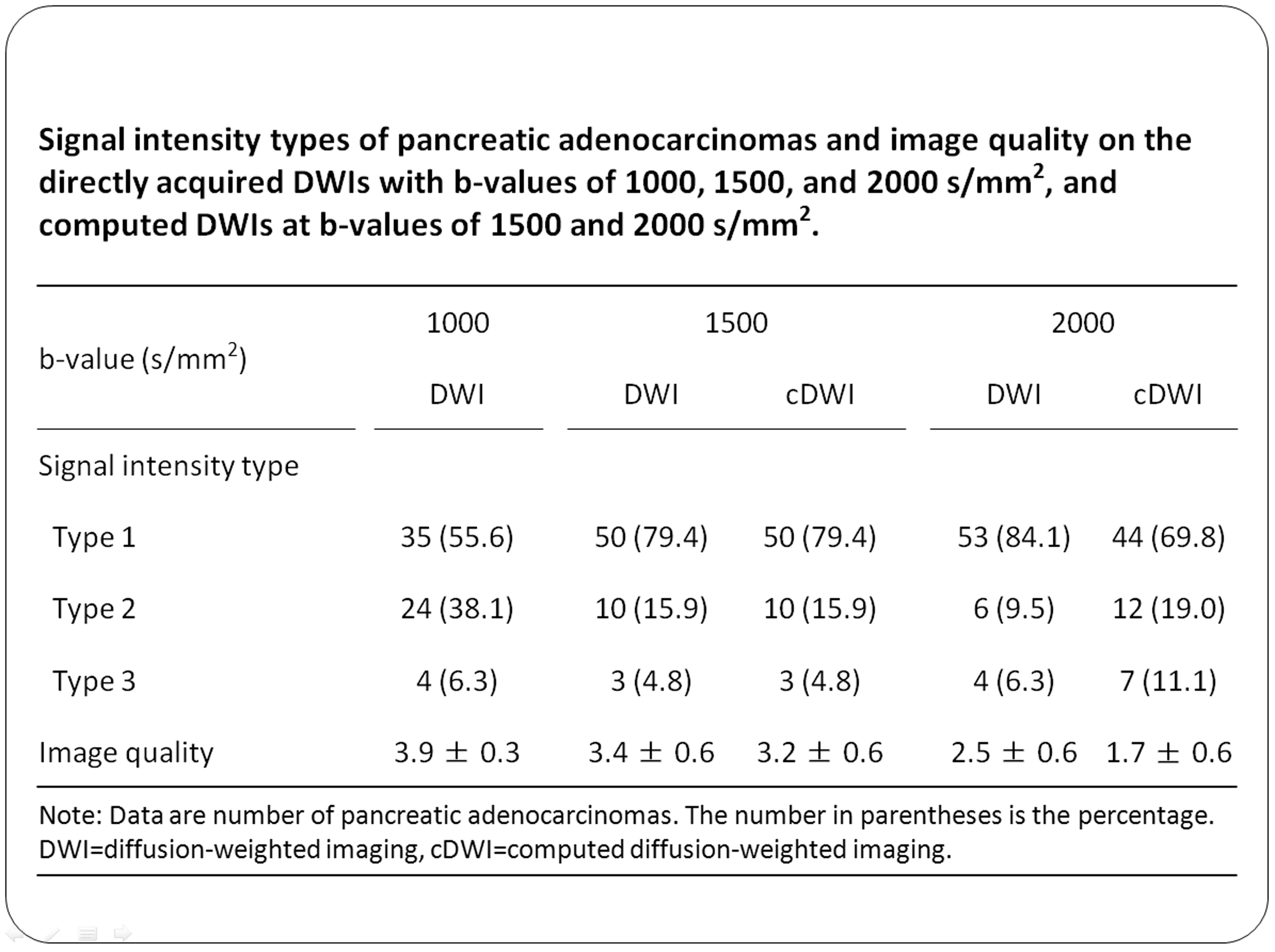
Sixty-three patients with histologically confirmed pancreatic adenocarcinoma underwent DWI at b-values of 0, 1000 (DWI1000), 1500 (DWI1500) and 2000 (DWI2000) s/mm2. From the DWIs at b-values of 0 and 1000 s/mm2, cDWIs with b-values of 1500 (cDWI1500) and 2000 s/mm2 (cDWI2000) were generated. We retrospectively assessed directly acquired DWI and cDWI findings of pancreatic adenocarcinomas (clear hyperintensity; hyperintensity with an unclear distal border; andResults
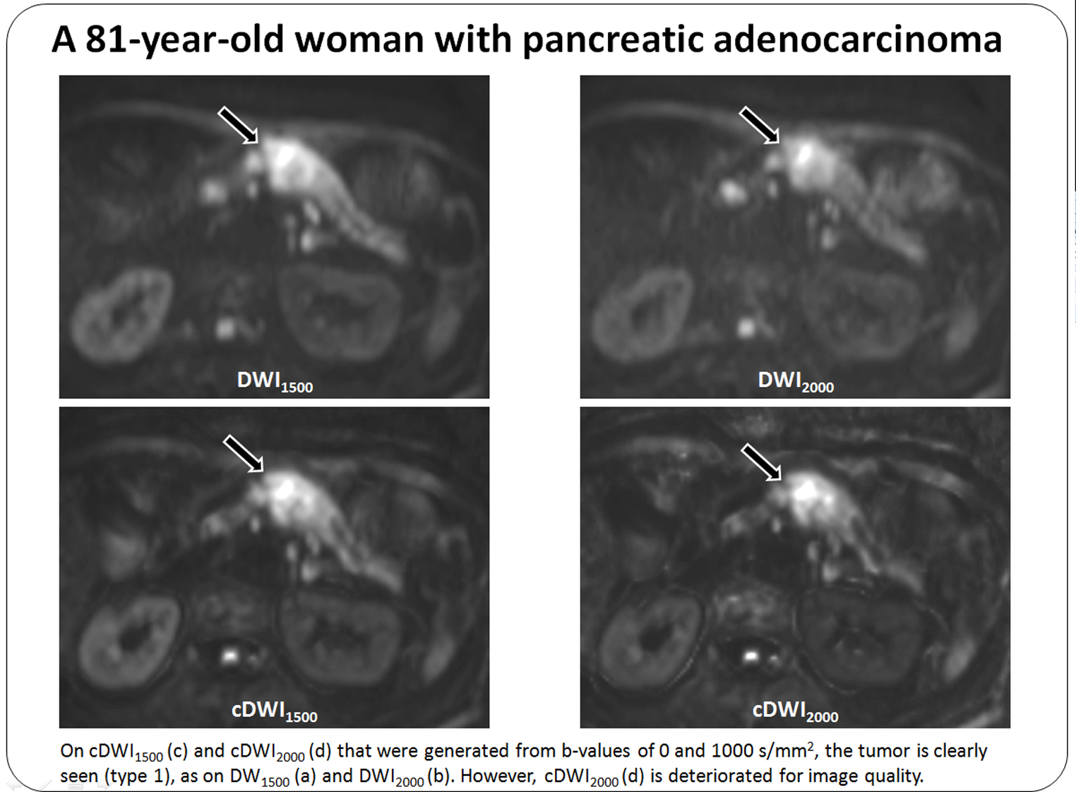
Clear hyperintense tumors were seen in 35 (55.6%) on DWI1000, 50 (79.4%) on DWI1500, 50 (79.4%) on cDWI1500, 53 (84.1%) on DWI2000 and 44 (69.8%) on cDWI2000 (Table 1). There was no significant difference in the incidence of clear hyperintense tumors between DWI1500 and cDWI1500 (P>0.999), but a lower incidence was seen on cDWI2000 than on DWI2000 (P = 0.028). Image quality was lower on cDWI than on DWI at b-values of 1500 s/mm2 (P = 0.002) and 2000 s/mm2 (P < 0.001) (Figure). There was no significant difference in the tumor to proximal CR between DWI and cDWI at b-values of 1500 s/mm2 (P = 0.063) and 2000 s/mm2 (P = 0.066), but the tumor to distal pancreas CR was significantly higher on cDWI than on DWI at b-values of 1500 s/mm2 (P = 0.004) and 2000 s/mm2 (P < 0.001).Discussion
In this study, clear hyperintense pancreatic adenocarcinomas were seen in increasing numbers of patients as the b-value increased in DWI: 35 (55.6%) on DWI1000, 50 (79.4%) on DWI1500 and 53 (84.1%) on DWI2000, which is supported by previous studies showing that pancreatic adenocarcinoma is not always clear on DWI at b-values ≤ 1000 s/mm2, and the tumor visualization improves when b-values increase (1,2). The incidence of clear hyperintense pancreatic adenocarcinoma on cDWI1500 was significantly higher than that ofConclusion
cDWI1500 can improve the delineation of pancreatic adenocarcinomas when compared with directly acquired DWI1000. Computed and directly acquired DWIs at a b-value of 1500 s/mm2 also produce comparable visualization of pancreatic adenocarcinomas. However, it is apparent that using cDWI may facilitate the incorporation of high b-value DWI into clinical interpretation schemes, since cDWI at higher b-values can be obtained in a shorter acquisition time and irrespective of the MRI system’s configuration.Acknowledgements
No acknowledgement found.References
1. Fukukura Y, Takumi K, Kamimura K, et al. Pancreatic adenocarcinoma: variability of diffusion-weighted MR imaging findings. Radiology 2012; 263:732-740.
2. Fukukura Y, Shindo T, Hakamada H et al. Diffusion-weighted MR imaging of the pancreas: optimizing b-value for visualization of pancreatic adenocarcinoma. Eur Radiol 2016; 26:3419-3427.
3. Blackledge MD, Leach MO, Collins DJ, Koh DM. Computed diffusion-weighted MR imaging may improve tumor detection. Radiology 2011; 261:573-581.
Figures