3536
White Matter Microstructural Changes in Healthy Aging: The Impact of Free Water Elimination on DTI Metrics1Rotman Research Institute, Baycrest Health Sciences, Toronto, ON, Canada, 2Brigham and Women’s Hospital, Harvard Medical School, Boston, MA, United States, 3Massachusetts General Hospital, Harvard Medical School, Boston, MA, United States, 4Medical Biophysics, University of Toronto, Toronto, ON, Canada
Synopsis
A Free Water Elimination (FWE) technique is applied to investigate changes in DTI parameters in the human white matter during healthy aging. Our results suggest that age-related changes in DTI metrics often reflect increased extracellular free water with age. DTI with FWE unmasks the effects of free water, revealing authentic tissue microstructure.
Purpose
The high anisotropy of diffusion within the white matter (WM) has made diffusion tensor imaging (DTI) a prominent mode of studying WM changes across the lifespan. A general trend has been an increase in mean diffusivity (MD) and decrease in fractional anistropy (FA) with age, suggesting overall decreased WM integrity [1]. This trend is most prevalent in frontal WM, supporting the concept of an "anterior-posterior" gradient of aging. However, it is well known that neural tissue degenerates with age [2], leading to increased partial voluming with extracellular free water. A free water elimination (FWE) technique has been proposed to obtain DTI parameters for a 'tissue compartment' which is separated from a 'free water compartment' [3]. This technique has been shown to be a promising tool for correcting for free water partial voluming in WM DTI parameters [4]. In this study, we apply the FWE technique to investigate authentic changes in WM microstructure during healthy aging.Methods
212 healthy subjects aged 39.1 to 91.7 years (62.0 + 11.2) were imaged on a Siemens Magnetom Trio system with a 12-channel head coil. Diffusion MRI (dMRI) with 60 directions was performed at b=700mm2/s following 10 b=0 volumes. Other parameters were TE=83ms, TR=7.98s, FOV=25.6cm, 2mmx2mmx2mm resolution, 176 slices. All dMRI data sets were corrected for motion and eddy currents by linearly registering each volume to the first b0 image [5] (eddy_correct in FSL [6]), and masked using FSL's Brain Extraction Toolbox (bet) [7]. FWE was performed as per Ref (3), resulting in DTI parameters specifically for "tissue" compartments, and a "free water map" providing the fraction of free water per voxel. Conventional, non-FWE DTI was also performed by fitting the single-tensor model. Voxelwise statistical analysis in the white matter was conducted using FSL's Tract-Based Spatial Statistics (TBSS) [8]. A relationship of DTI parameters with age was defined by p<0.05 with correction for multiple comparisons and controlling for gender.
Results
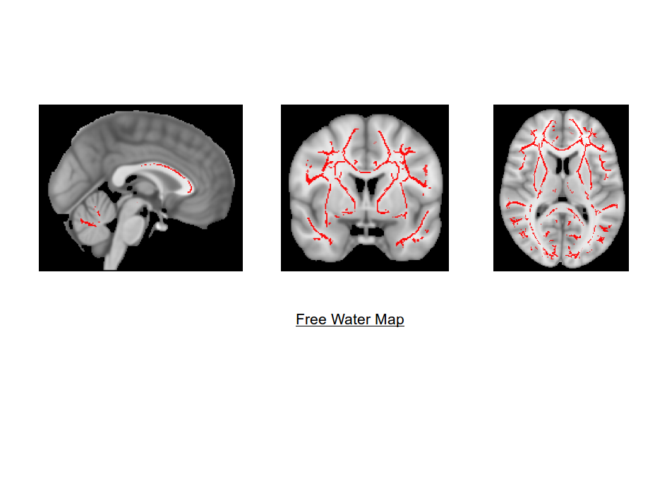
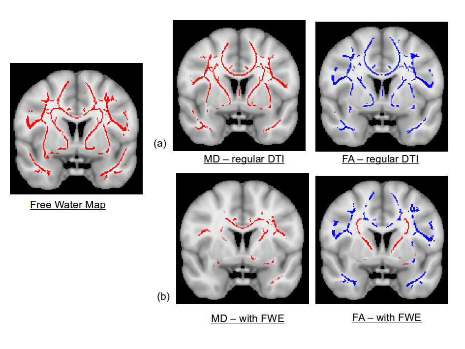
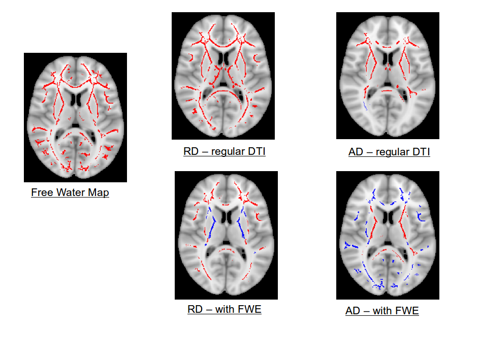
TBSS results for the DTI model show increases in MD and decreases in FA with age (e.g. Figure 2a), as well as RD increases throughout the WM, most prevalent in anterior regions. Correlations of age and AD are less persistent, with regions of both increased and decreased AD with age. This is all consistent with the literature [1]. Free water maps reveal increased free water with age throughout the white matter (Figure 1). This increase with age in free water largely overlaps with the FA, MD and RD changes with age. After free water elimination, many perceived changes in DTI parameters with age vanish (Figure 2b). FWE still shows regions of increased RD with age, although less numerous than in regular DTI. In the superior corona radiata and internal capsule, FWE reveals decreased RD with age (Figure 3), which contrasts the known prevalency of RD increase from regular DTI. Additionally, several areas which show increased AD with age in DTI are shown to have decreased AD with age after FWE.
Discussion
The DTI results are consistent with the literature [1]. The structure of the free water maps is also consistent with the previously reported anterior-posterior gradient of aging. While regular DTI shows increased AD with age in much of the WM, FWE unmasks several of these regions to show decreased AD with age, possibly due to age-related accumulation of intracellular debris. Various regions still show increased RD with age after the FWE, suggesting an authentic decrease in WM integrity in these regions. Moreover, the RD decrease with age shown by FWE in the superior corona radiata and internal capsule is accompanied by an AD increase (and thus an FA increase). This may be due to an asymmetrical degeneration of crossing fibers with age.
It should be noted that variations in white matter volume across the lifespan can lead to potential misalignments when registering all skeletons to MNI space, and furthermore the native data were not corrected for EPI artifacts. Additionally, the FWE does not account for partial voluming of gray matter and WM which may vary between subjects. Efforts to additionally study WM changes in native space are underway.
Conclusion
Free water maps reveal an increase in extracellular free water in the white matter with age, highlighting the importance of FWE when using DTI to investigate WM microstructure during aging. FWE reduces the number of regions of age-associated microstructural change, suggesting that partial-volume effects with free water are significant in regular DTI processing. Controlling for this can put better focus on specific regions that may have authentic microstructural age-related changes.Acknowledgements
We thank the Canadian Institutes of Health Research (CIHR) and the National Institutes of Health (NIH) for financial support.References
1. Madden EJ, et al. Diffusion tensor imaging of ceberal white matter integrity in cognitive aging. Biochimica et Biophysica Acta, 1822(3): 386-400, 2012
2. Peters R. Ageing and the brain. Postgrad Med J, 82(964): 84-88, 2006
3. Pasternak O, Sochen N, Gur Y, Intrator N, Assaf Y. Free water elimination and mapping from diffusion MRI. Magn Reson Med 62(3):717-730, 2009.
4. Metzler-Baddeley C, O'Sullivan MJ, Bells S, Pasternak O, Jones DK. How and how not to correct for CSF-contamination in diffusion MRI. Neuroimage 59(2): 1394-1403, 2012
5. Jenkinson M, Smith S. A global optimisation method for robust affine registration of brain images. Med Image Anal, 5(2): 143-156, 2001
6. Smith SM, et al. Advances in functional and structural MR image analysis and implementation as FSL. Neuroimage, 23 Suppl 1: S208-219, 2004
7. Smith SM. Fast robust automated brain extraction. Hum Brain Mapp, 17(3): 143-155, 2002
8. Smith SM, et al. Tract-based spatial statistics: voxelwise analysis of multi-subject diffusion data. Neuroimage, 31(4): 1487-1505, 2006
Figures