3526
SHANK-3 gene mutation results in comprehensive white matter damage in children: a DTI-TBSS study1Children's Hospital of Fudan University, Shanghai, People's Republic of China, 2Laboratory of Biomedical Imaging and Signal Processing, The University of Hong Kong, HongKong, Hong Kong
Synopsis
Autism spectrum disorder (ASD) is classified as a neuro-developmental disease with a dramatically increasing prevalence from 4 in 10000 to recently 1 in 68 children. SHANK-3 proteins are multidomain scaffold proteins of the postsynaptic density and also play a role in synapse formation and dendritic spine maturation. Recent human genetic studies suggest the potential association between molecular defects of SHANK-3 and ASD. DTI imaging and TBSS analysis was applied to study how SHANK-3 gene mutation results in severe microstructure of white matter. Results showed significant damage in SHANK-3 group but no positive findings between ASD and typical development controls. These results calls for attention to re-examine the previous neuroimaging studies of ASD or other neuro-developmental diseases where the positive correlations could be contaminated with unexplored genetic mutation influence.
Target audience
Scientists and clinicians specializing in neuro-developmental disease, autism spectrum disorder, or gene-related cognitive deficits.Purpose
Autism spectrum disorder (ASD) is classified as a neuro-developmental disease with a dramatically increasing prevalence from 4 in 10000 to recently 1 in 68 children1-3. Genetic factors are believed to contribute to the underlying pathophysiological mechanisms4-6. SHANK-3 proteins are multidomain scaffold proteins of the postsynaptic density and also play a role in synapse formation and dendritic spine maturation7-10. Recent human genetic studies suggest the potential association between molecular defects of SHANK-3 and ASD8-10. DTI, as an invasive examination tool, has been widely applied to developing brains to study the white matter changes11. Numerous studies reported the reduced FA indicating increased WM microstructural disorganization in autistic population12,13. However, all those studies only evaluated the severity of ASD by relating to clinical manifestations or observation scales but none has categorized ASD based on genotypes. In this work, we aimed to document the relationship between specific white matter changes and genetic mutations. We tested the gene mutation for each of the diagnosed ASD children by dividing the ASD group into two subgroups: one without obvious gene deficits, and the other with SHANK-3 gene mutation.Methods
56 children (2-9 yrs) were included in the present study. They included 30 ASD subjects who were selected according to the criteria of DSM-V with a cut-off score of ADOS. MLPA technique was applied to all of the ASD patients to identify the SHANK-3 mutation and eliminate other apparent genetic deficits. Among them, 8 subjects were identified with SHANK-3 mutation while 22 subjects did not present genetic deficits. Further, 26 subjects with typical development (TD) and no neurologic or degenerative diseases were included. They underwent MRI scan because of febrile convulsion, unexplained headache, unexplained dizziness or mild trauma. MR scans were obtained on a GE 3.0 Tesla Discovery MR750 system (GE Medical Systems, Milwaukee, WI) with a 32-channel head coil. DTI datasets were acquired using an echo-planar imaging sequence with TR 4600ms, TE 88 ms, 15 directions uniformly distributed in three-dimensional space, 15 B-factors 0 and 1000 s/mm2, axial slices covering the whole brain. All DTI datasets were first corrected for eddy current distortions using EDDY embedded in FSL (FMRIB Software Library). Then DTIFIT fitted a diffusion tensor model at each voxel to generate FA, AD, RD and ADC maps. The TBSS analysis was again performed using the FSL. All FA images were aligned to form the target image. This target image was then affine-aligned into MNI152 standard space, and every image was transformed into 1x1x1mm MNI152 space by combining the nonlinear transform to the target FA image with the affine transform from that target to MNI152 space. Normalized FA maps were visually assessed to ensure good normalization quality. Tract-based spatial statistics were then tested with FA maps to create the "skeleton", which represents the center of all fiber bundles in common to all subjects, using a threshold of FA > 0.2. Each subject’s aligned FA data were then projected onto this skeleton, and the resulting data were fed into voxel-wise cross-subject statistics. Nonparametric statistical analysis was performed using FSL based on permutation analysis applied to the general linear model (5000 permutations). Resulting statistical maps were thresholded at P < 0.001 corrected for multiple comparisons at a cluster level using the threshold-free cluster enhancement (TFCE) approach.Results
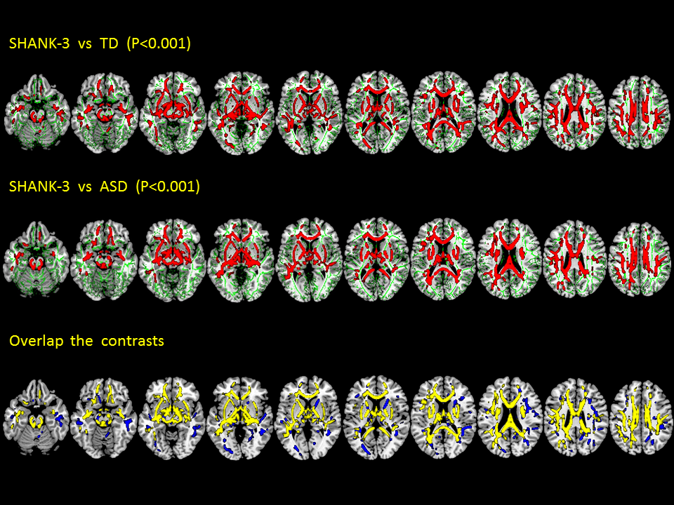
TBSS post-statistical analysis between SHANK-3 group (n=8) and typical development (TD; n=22) control, as well as the results between SHANK-3 group and entire ASD group (n=30), clearly showed significant FA decreases on the comprehensive white matter networks (see the attached Figure). The main affected fibers are (P<0.001): (both left and right sides) cingulum, corpus callosum, cortico spinal, cortico cerebellum, fornix, inferior longitudinal fasciculus, inferior occipital frontal fasciculus, internal capsule. However, interestingly, even on p<0.05 level. We found no significantly different FA values in any white matter regions between ASD group and TD group.Conclusions
This work clearly demonstrates that SHANK-3 gene mutation results in severe microstructural damage of all major white matter networks. The negative finding between ASD group and TD group calls for attention to re-examine the previous neuroimaging studies of ASD or other neuro-developmental diseases where the positive correlations could be contaminated with unexplored genetic mutation influence. Therefore, genotype should also be included as one of the grouping criteria in the future neuroimaging studies to reduce subject heterogeneity.Acknowledgements
No acknowledgement found.References
1. “Autism spectrum disorder fact sheet” .DSM5.org. American Psychiatric Publishing. 2013.
2. American Psychiatric Association. Diagnostic and statistical manual of mental disorders.5th edition.Washington, DC: American Psychiatric Association; 2013.
3. Centers for Disease Control and Prevention. Prevalence of autism spectrum disorder among children aged 8 years—autism and developmental disabilities monitoring network,11Sites, United States, 2010. MMWR Surveill Summ 2014;63(2):1–21.
4. Elsabbagh M, Divan G, Koh YJ, Kim YS, Kauchali S, Marcin C, Montiel-Nava C, Patel V, Paula CS, Wang C, Yasamy MT, Fombonne E (2012) Global prevalence of autism and other pervasive developmental disorders. Autism Res 5:160-179.
5. Jeste SS, Geschwind DH (2014). Disentangling the heterogeneity of autism spectrum disorder through genetic findings. Nat Rev Neurol 10: 74-81.
6. Persico AM, Napolioni V (2013) Autism genetics. Behav Brain Res. doi: 10.1016/j.bbr.2013.06.012 7. Boeckers TM, Bockmann J, Kreutz MR, Gundelfinger ED (2002). "ProSAP/Shank proteins - a family of higher order organizing molecules of the postsynaptic density with an emerging role in human neurological disease". J. Neurochem. 81 (5): 903–10.
8. Nemirovsky S.I., Córdoba M., Zaiat J.J., Completa S.P., Vega P.A., González-Morón D., Medina N.M., Fabbro M., Romero S., Brun B., Revale S., Ogara M.F., Pecci A., Marti M., Vazquez M., Turjanski A., Kauffman M.A. Whole genome sequencing reveals a de novo SHANK3 mutation in familial autism spectrum disorder. PLoS One, [Online] 2015;10
9. Jiang Y-H., Ehlers M.D. Modeling autism by SHANK gene mutations in mice. Neuron. 2013;78:8–27.
10. Shigeo U, Chikako W. Novel Therapeutic Approach for Autism Spectrum Disorder: Focus on SHANK3:[J]. Dna Research An International Journal for Rapid Publication of Reports on Genes & Genomes, 2015, 13(999):786-792.
11. Jou RJ, Jackowski AP, Papademetris X, Rajeevan N, Staib LH, Volkmar FR. Diffusion tensor imaging in autism spectrum disorders: Preliminary evidence of abnormal neural connectivity. Aust N Z J Psychiatry. 2011; 45(2):153–162.
12. Sundaram SK, Kumar A, Makki MI, Behen ME, Chugani HT, Chugani DC. Diffusion tensor imaging of frontal lobe in autism spectrum disorder. Cereb Cortex. 2008; 18(11):2659–2665. Shukla DK, Keehn B, Smylie DM, Müller R. Microstructural abnormalities of short-distance white matter tracts in autism spectrum disorder. Neuropsychologia. 2011; 49(5):1378–1382.
13. Shukla DK, Keehn B, Smylie DM, Müller R. Microstructural abnormalities of short-distance white matter tracts in autism spectrum disorder. Neuropsychologia. 2011; 49(5):1378–1382.
Figures