3515
Feasibility of very high b-value diffusion imaging using a clinical scanner.1Center for Magnetic Resonance Research, Minneapolis, MN, United States
Synopsis
The use of locally low-rank matrix approximation methods for noise variance reduction provides a powerful tool for diffusion MRI to increase the quality of very high b-value acquisitions. Using a clinical system, we demonstrate the feasibility of multi-shell high angular resolution diffusion imaging with b-value up to 10,000s/mm2.
Purpose
In diffusion MRI (dMRI), the choice of b values and gradients directions (i.e. q-space sampling) is driven by the need to achieve relatively high spatial resolution, while covering q-space as optimally as possible to minimize the angular uncertainty of fiber orientations [1]. Protocol optimization based on noise-averaging are biased towards lower b-values. Reconstruction methods leveraging the high dimensionality of (relatively long) diffusion acquisitions to minimize noise variance offer a powerful tool to enhance the signal-to-noise ratio of current state-of-the-art imaging protocols [1]. We demonstrate the benefits of such approaches by assessing their ability to identify voxels with fiber crossings (up to three fibers) and minimize their associated uncertainty. This is achieved using data from a clinical scanner with b-value up to 10,000 s/mm2.Methods
Data was acquired on a 3T MAGNETOM Prisma system (Siemens), equipped with 80mT/m gradient set, in accordance with IRB approval from the university of Minnesota. Diffusion encoding was performed using four shells with b-values 1000, 3000, 6000 and 10000 s/mm2 , each with 128 directions and 15 additional b=0 volumes, in AP and PA phase encoding directions (for a total of 290 volumes). For whole brain diffusion acquisition the C2P[2] used for the human connectome[3] and life-span project was used, with MB factor=4, voxel size=2mm isotropic, TE/TR=105ms/3000ms matched for all b-values (defined by the b=10000 s/mm2 shell)
Noise variance reduction was performed on the complex-valued SENSE1 combined images, using a parameter free locally low-rank (LLR) denoising technique calibrated to the spatio-temporal incoherence of thermal noise. The Casorati matrix used for noise-removal was applied (slice by slice) to spatial patches with size 35x35, and across all 1160 volumes simultaneously.
Prior to analysis, pre-processing using eddy and topup was applied [1]. After segmentation of the white matter and identification of the corona radiata by nonlinear registration of the Johns Hopkins University white matter atlas [4], the proportion of voxels with two and up to three fibers was estimated. For each fiber, orientation’s uncertainty was obtained from FSL bedpostX [5].
Results
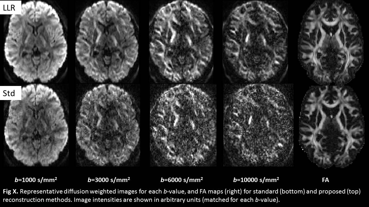
The reconstruction provided by the vendor software, using the C2P, was compared with an offline reconstruction based on a parameter free LLR soft thresholding denoising method. Representative images after distortions correction (but before averaging of AP and PA volumes) for each b-value, with and without noise-variance reduction, are shown in Figure 1. For each b-value, an arbitrary scaling is applied (consistent across reconstruction methods), since the difference in signal between the low and high b-values is an order of magnitude. The column on the right shows the FA calculated from all volumes.
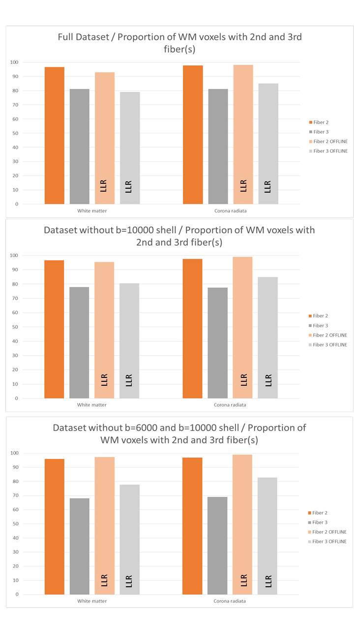
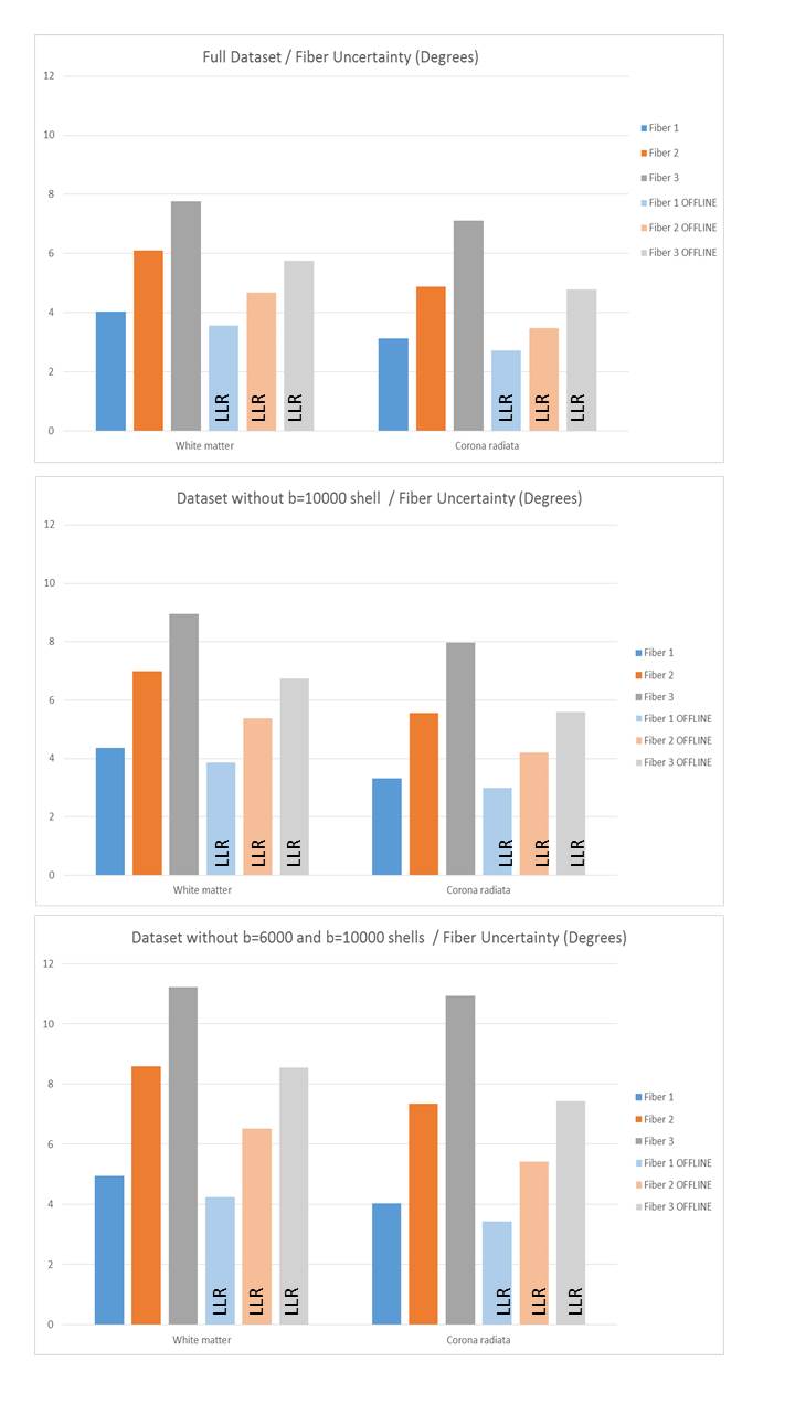
To assess the additional information provided by high b-values, depending on the reconstruction method employed, two subsets of the full dataset were also analyzed: One without the b=10000s/mm2 shell, and one without the two highest b-values (10000 and 6000s/mm2). The results for the proportion of white matter voxels with two or three fibers are shown in Figure 2, as well as uncertainty in Figure 3.
Discussion
The proportions of white matter voxels with two and up to three fibers are relatively similar for both reconstruction methods. However, the proposed reconstruction helps recovers more fibers (results close to those using the full dataset) with down-sampled datasets. For the fiber uncertainty, the denoised data consistently exhibit lower angular uncertainty, both for the full dataset and for subsets hereof. For the online data, doubling the acquisition from two shells to 4 shells improves the uncertainty approximately by about 1 to 3 degree. For the dataset with denoising, the third fiber uncertainty is reduced by 2 degrees relative to the full on-line data, whereas the changes for the 1st and 2nd fiber is approximately reduced ½ and 1 degree respectively.Conclusion
The reduction in angular uncertainty for the third fiber obtained with denoising versus the raw data is a factor of 2. The use of a parameter free LLR denoising technique, which does not exploit spatial features, is desirable for diffusion imaging, since the changes through diffusion volumes vary both spatially and angularly. The use of very high b-values is normally restricted due to associated long echo times, and subsequent lower SNR. With the proposed LLR technique, higher b-values (potentially even higher than 10000s/mm2) can be incorporated into diffusion protocols, application of the denoising techniques should be factored in a-priori when selecting how to acquire large diffusion acquisitions.Acknowledgements
P41 EB015894References
[1] Sotiropoulos SN et al. Advances in diffusion MRI acquisition and processing in the Human Connectome Project. Neuroimage. 2013 Oct 15;80:125-43. doi: 10.1016/j.neuroimage.2013.05.057.
[2] http://www.cmrr.umn.edu/multiband
[3] Ugurbil K, Pushing spatial and temporal resolution for functional and diffusion MRI in the Human Connectome Project. Neuroimage. 2013 Oct 15;80:80-104. doi: 10.1016/j.neuroimage.2013.05.012. Epub 2013 May 21.
[4 ]Wakana S. Reproducibility of quantitative tractography methods applied to cerebral white matter NeuroImage, 36 (2007), pp. 630–644
[5] Jenkinson M. FSL, Neuroimage. 2012 Aug 15;62(2):782-90
Figures