3513
Quantification of Navigator Requirements for Multi-shot Diffusion Weighted Imaging1Bioengineering, University of Illinois at Urbana-Champaign, Urbana, IL, United States
Synopsis
The ability to use multiple shots in diffusion weighted imaging has enabled acquisitions with higher resolutions and higher SNRs. The methods currently being used to achieve higher resolutions rely on navigator information to correct for mismatches in coherent motion during the diffusion encoding between shots. The resolution of the navigator is a critical factor, too low and the corrections are incomplete, too high and there is a significant time or echo time penalty. This work presents an analysis for determining the resolution requirements for navigation. This work found that using a navigator resolution of 6 mm produces reliable results and allows shorter acquisitions to be used.
Introduction
Diffusion weighted imaging (DWI) provides contrast to tissue microstructure. DWI had been traditionally limited to lower spatial resolutions due to the need to encode all of k-space in a single shot due to motion induced phase.1 Recently there has been a move towards acquisitions that use navigator information to reduce artifacts of motion induced phase error, allowing higher resolutions to be achieved.2-4
Many multi-shot diffusion protocols use a large navigator, sometimes requiring the use of a second echo and resulting in a reduction of scan-time efficiency. The phase information in the navigators are typically lower resolution, and do not require sampling of high frequency k-space information in order to accurately model the phase errors, however, the requirements for the navigator have not been fully analyzed. In this work, the impact of the navigator resolution on the SNR of a reconstructed image will be presented, allowing for the potential of faster acquisitions by shortening navigators or moving them to more efficient locations.
Methods
All experiments were performed on a Siemens Trio scanner using a gradient strength of 26 mT/m for diffusion encoding. For all data shown, diffusion encoding was in the superior/inferior direction as previous work has shown it is the most susceptible to motion induced phase errors.5 All images used spiral trajectories with an iterative reconstruction to form an image with a 120 matrix size.6 For multi-shot data, images were reconstruction using the CG phase method.7
First a healthy subject was scanned using a single shot (R=2) sequence with cardiac gating. A single axial slice was acquired at the level of the midbrain with 1 image acquired at every cardiac cycle. This area is known to have high nonlinear motion induced phase error.
Second, 3 volunteers were scanned using a free breathing 2 shot spiral scan with 40 2 mm slices acquired centered on the midbrain. The scan was repeated 25 times. Navigator images being obtained by reconstructing images from each shot separately (R=2). Different spatial resolution navigators were formed by discarding the appropriate high frequency k-space data in order to reconstruct navigators at different resolutions. The temporal SNR of the reconstructed images over the 25 repeats was calculated for each of the different resolution navigator cases.
Results
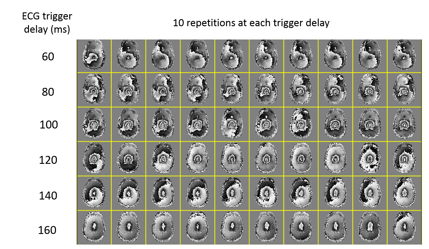
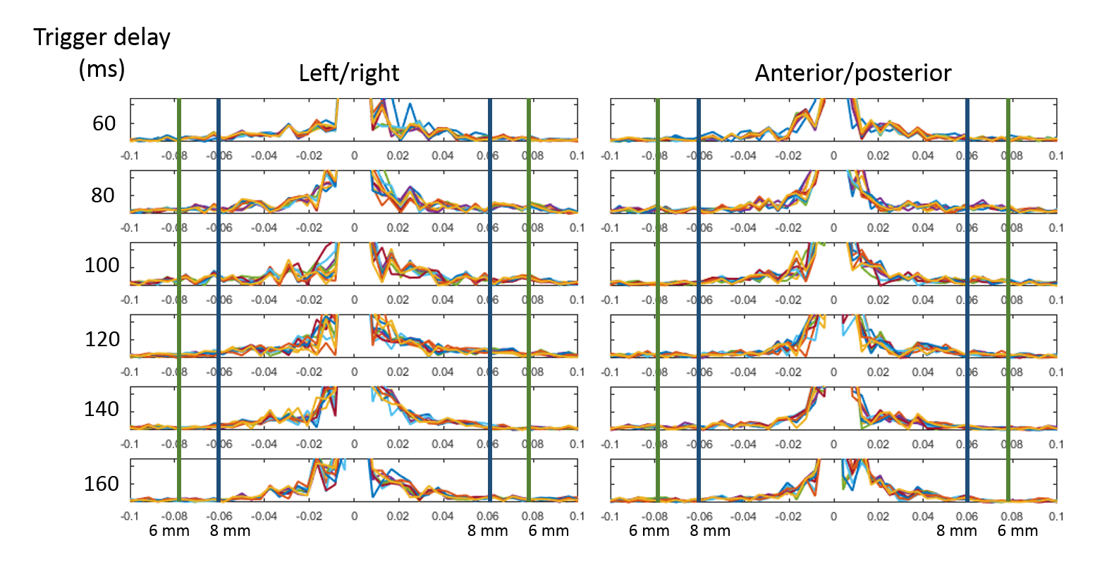
Figure 1 shows the phase images from a subject over 10 repetitions of the same measurement at different trigger delays. The profile Fourier transform of the phase images from figure 1 are shown in Figure 2 to indicate the regions of k-space that have significant energy.
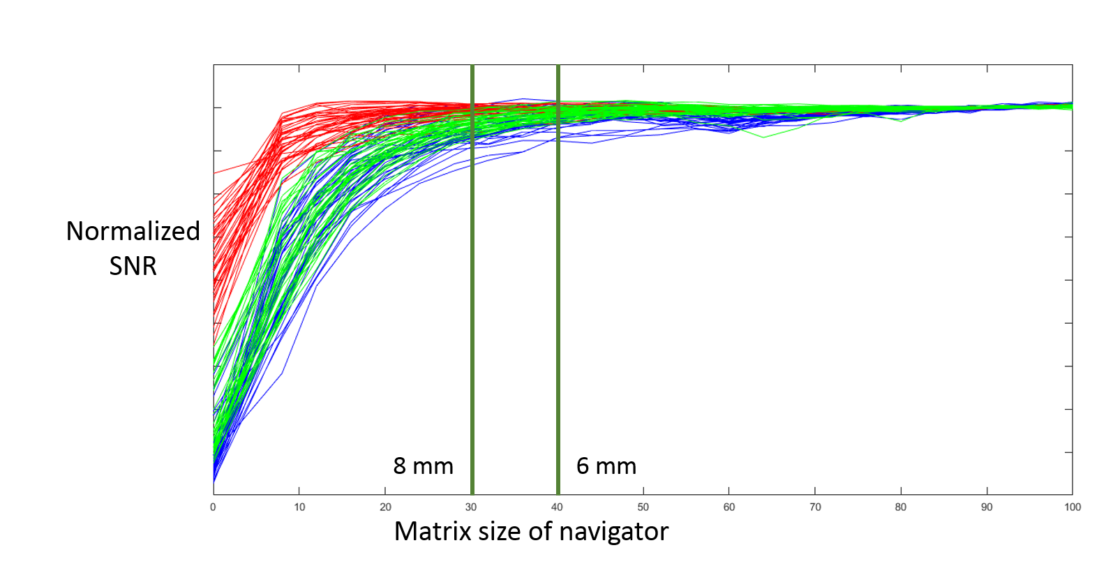
To demonstrate the impact of phase errors on the image reconstruction, Figure 3 shows the resulting temporal SNR of the series with different navigator resolutions. The colors show different subjects and the multiple lines are different slices within the imaging volume.
Discussion
From figure 1, the peak motion occurs at an ECG trigger delay of 100 ms. The motion appears to dissipate fairly rapidly suggesting that only a small part of the cardiac cycle may be impacted by this cardiac motion. Examining the spatial frequencies in the data in Figure 2 most of the phase information can be captured with an 8 mm resolution navigator. However, during peak pulsation in the cardiac cycle, a higher resolution navigator may be needed for accurate image reconstruction.
As the navigator resolution increases in Figure 3 the SNR reaches a plateau. The start of the plateau represents the resolution where the increase in navigator resolution does not further improve the reconstruction. This happens at a navigator resolution of 6 mm for most of the subjects. Using a navigator of lower resolution resulted in a loss of SNR. While the graph shows the shape of the loss in SNR. In practicality, this will depend on the number of shots used and the k-space sampling, so the emphasis should be placed on operating at a navigator resolution in the plateau region.
These results represent progress towards quantification of navigator sampling requirements, however it is important to realize that the phase errors will also depend on the size, shape, and direction of the diffusion encoding gradients. Using a navigator resolution of 6 mm enables us to move the navigator to a spiral-in location prior to the echo without extending the echo time.2
Conclusion
Acquiring navigators at lower resolutions still provides enough information for corrected for motion induced phase errors. This allows less of k-space to be sampled for navigators enabling shorter acquisition times and improved SNR efficiency. This work represents a step towards better understanding phase errors in DWI and design of navigators for improved acquisitions.Acknowledgements
No acknowledgement found.References
1. Anderson, A.W. and J.C. Gore, Analysis and correction of motion artifacts in diffusion weighted imaging. Magn Reson Med, 1994. 32(3): p. 379-87.
2. Holtrop, J.L. and B.P. Sutton, High spatial resolution diffusion weighted imaging on clinical 3 T MRI scanners using multislab spiral acquisitions. Journal of Medical Imaging, 2016. 3(2): p. 023501-023501.
3. Setsompop, K., et al., Pushing the limits of in vivo diffusion MRI for the Human Connectome Project. Neuroimage, 2013. 80: p. 220-33.
4. Engstrom, M. and S. Skare, Diffusion-weighted 3D multislab echo planar imaging for high signal-to-noise ratio efficiency and isotropic image resolution. Magn Reson Med, 2013. 70(6): p. 1507-14.
5. Wirestam, R., et al., Theoretical and experimental evaluation of phase-dispersion effects caused by brain motion in diffusion and perfusion MR imaging. Jmri-Journal of Magnetic Resonance Imaging, 1996. 6(2): p. 348-355.
6. Sutton, B.P., D.C. Noll, and J.A. Fessler, Fast, iterative image reconstruction for MRI in the presence of field inhomogeneities. IEEE Trans Med Imaging, 2003. 22(2): p. 178-88.
7. Liu, C., M.E. Moseley, and R. Bammer, Simultaneous phase correction and SENSE reconstruction for navigated multi-shot DWI with non-cartesian k-space sampling. Magn Reson Med, 2005. 54(6): p. 1412-22.
Figures