3506
High Resolution Cervical Spine Single-Shot DTI with Distortion Correction Using Modified Field-mapping Method1Center for Biomedical Imaging Research, Department of Biomedical Engineering, School of Medicine, Tsinghua University, Beijing, People's Republic of China
Synopsis
Single-shot echo planar imaging (EPI) DTI (SS-DTI) has been commonly used in clinical cervical spine (C-spine) MR scans to get functional and pathologic information as it’s fast and straightforward, but high resolution SS-DTI image suffers from severe geometric distortion. Field mapping method is a classical and simple distortion correction technique, but its performance in C-spine SS-DTI is limited. We used echo planar spectroscopic imaging (EPSI) sequence to acquire the field-map, and modified the conventional field-map post-processing method to promote the correction efficacy. The results show that the proposed method is effective and fast in C-spine SS-DTI distortion correction.
Purpose
Diffusion tensor imaging (DTI) plays a significant role in clinical cervical spine (C-spine) MR scans as it can reveal functional and pathologic information of the spinal cord through quantitative diffusion metrics. The conventional single-shot EPI DTI (SS-DTI) is fast and straightforward, but geometric distortion in high resolution images is severe, due to the long readout window and strong field inhomogeneity. Multi-shot EPI DTI (MS-DTI) can reduce distortion, but is time consuming and needs additional phase variation correction. Thus SS-DTI is still commonly used in clinical C-spine MR scans at present. The conventional field mapping method1 can provide necessary information for distortion correction. However, its performance in C-spine DTI is limited due to the complicated magnetic field environment and relatively lower SNR. This study aims to propose an effective and fast distortion correction method for C-spine SS-DTI, based on a modified field-map acquisition and post-processing pipeline.Methods
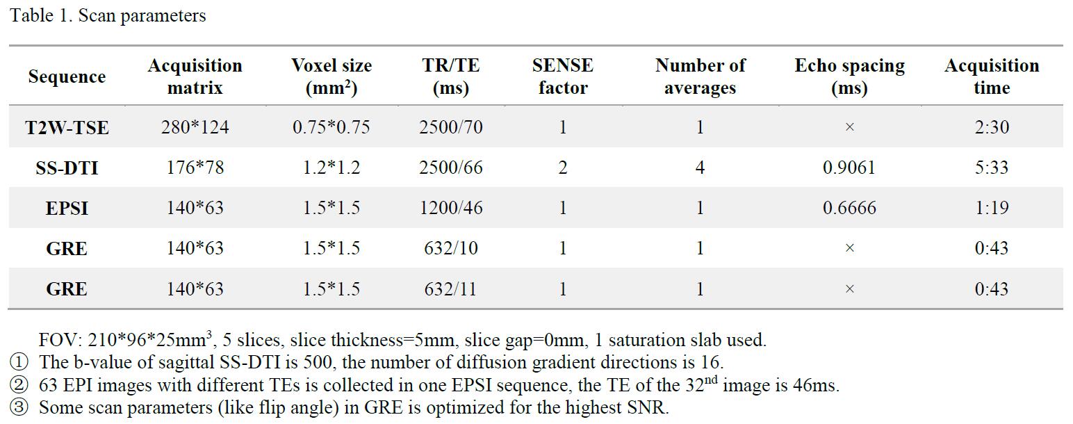
Data Acquisition All scans were performed on a Philips 3.0T Achieva TX MR scanner (Philips Healthcare, Best, The Netherlands) using a sixteen-channel SENSE neurovascular coil. Data were acquired from 5 healthy volunteers. The field inhomogeneity map was acquired using an echo planar spectroscopic imaging (EPSI) sequence2 (Fig. 1) in the sagittal view, which can provide multiple low resolution distortion-free spin-echo (SE) EPI images with equally distributed TEs. For comparison, Gradient echo (GRE) two different TEs was scanned as the conventional field mapping method. The in-plane resolution of sagittal SS-DTI was 1.2*1.2mm2. T2W-TSE was acquired as an anatomical reference to evaluate the correction efficacy. The detailed scan parameters were listed in Table 1.
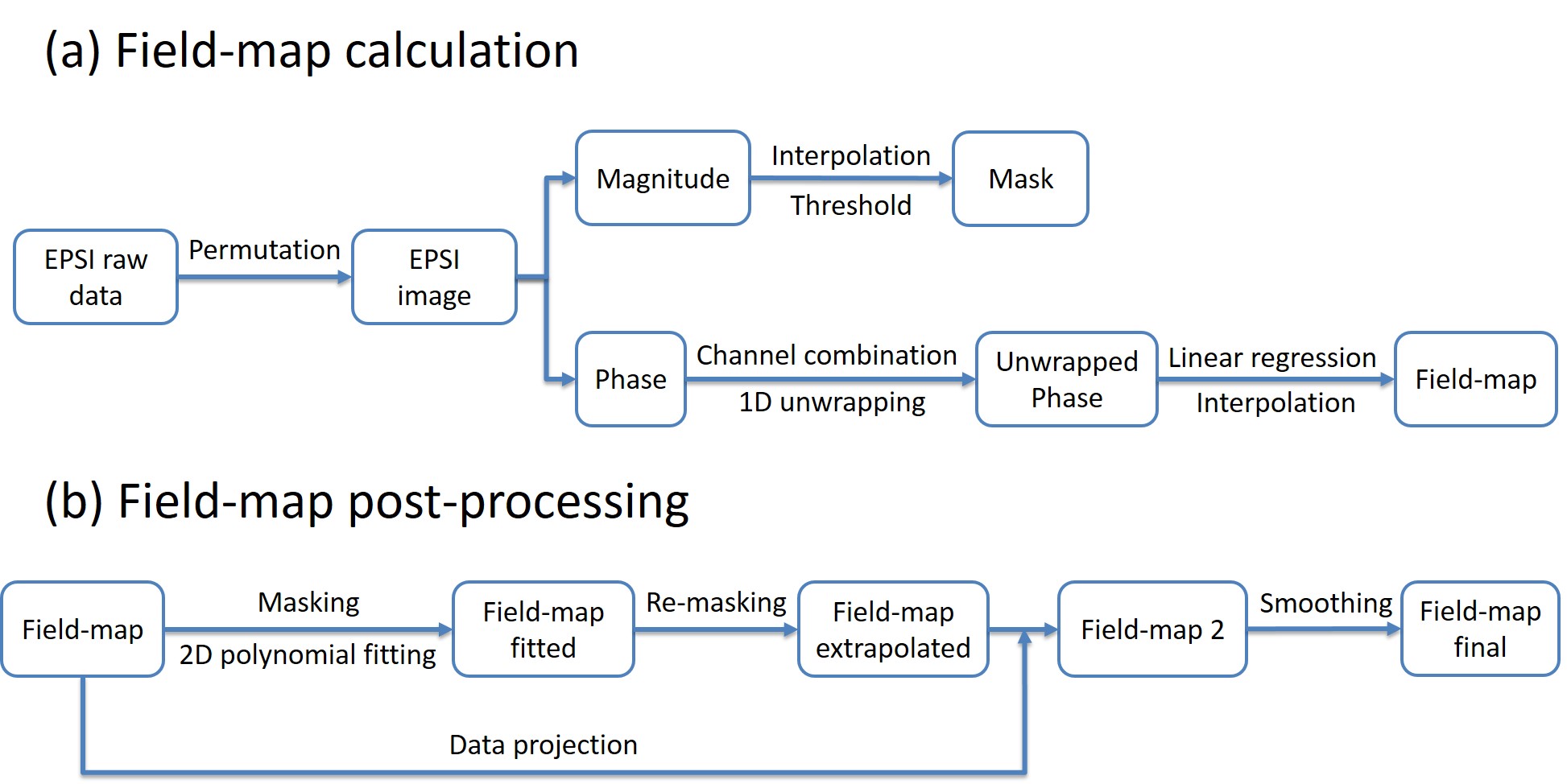
Data Processing The EPSI data were reconstructed after a
simple permutation of the data. The phase images from all channels were combined
using SENSE before the field-map calculation. Then one-dimensional phase
unwrapping of the phase images along the TE dimension was implemented, followed
by pixel-wise linear regression of the unwrapped phase images as a function of
TE to derive the field-maps. The field-map was then extrapolated by several
pixels (5 in this experiment) by 2D 3-order polynomial fitting and re-masking.
Then the original field-map was projected onto the extrapolation result, and
the whole field-map was slightly smoothed. The complete procedure of the field-map
processing was shown in Fig. 2. Field-map from GRE was also calculated and
processed with the same operations for comparison. Image distortion correction
was then implemented using the conventional coordinates searching and linear
interpolation algorithm1 in the image
domain.
Results and Discussion
As is shown in Table 1, EPSI collects 63 EPI images in one sequence to calculate a field-map, while GRE gets only 2 images with longer scan time. 1D phase unwrapping along the TE dimension is more robust than traditional 2D in-plane phase unwrapping under our imaging condition2, and the linear regression algorithm can reduce the influence of noise. Furthermore, EPSI is a SE-based sequence. So EPSI can provide more accurate field-map with higher SNR than GRE in structure with complex magnetic field environment like the C-spine.
Fig. 3 shows the correction results for relatively mild distortion. In uncorrected SS-DTI images (h, o) we can observe the variation of spinal cord curve radian. EPSI field-map (b) can correct the distortion (i, p), but there are some residual artifacts (yellow arrowheads) as the field inhomogeneity right outside the spinal cord boundary is unknown. Field extrapolation (c) can reduce these artifacts (j, q), but the complex magnetic field distribution in spinal cord can’t be estimated accurately by polynomial fitting. This may reduce the effect of correction (blue arrowheads), so a data projection operation (d) is necessary. Smoothing the field-map (e) can reduce artifacts (green arrowheads) resulted from the field discontinuity, and generate relatively satisfied correction results (l, s). On the other hand, GRE field-map (f) is noisier than the EPSI field-map (b-e) under the same window levels, and leads to correction results with strong artifacts (m, t). GRE field-map after the same processing (g) still cause some artifacts (blue arrowheads).
Fig. 4 shows the distortion correction results using EPSI field-map with complete post-processing in another case. After distortion correction, the sawtooth boundary of spinal cord (yellow boxes) is well restored, and the cervical spinal cord curve (yellow arrowheads) after correction is consistent with the TSE images.
Conclusion
In this study, a distortion correction method based on EPSI field-map acquisition and modified post-processing was proposed. The results demonstrate that this method has the potential in C-spine SS-DTI distortion correction, which may improve the image quality of high resolution SS-DTI. By using this strategy, it is possible to achieve fast and reliable C-spine diffusion imaging.Acknowledgements
No acknowledgement found.References
[1] Peter Jezzard, and Robert S. Balaban. "Correction for geometric distortion in echo planar images from B0 field variations." Magnetic resonance in medicine34.1 (1995): 65-73.
[2] Chen Bin, Hua Guo, and Allen W. Song. "Correction for direction-dependent distortions in diffusion tensor imaging using matched magnetic field maps."Neuroimage 30.1 (2006): 121-129.
Figures