3464
The histological validation analysis of diffusional kurtosis imaging with a cleared mouse brain.1Radiology, Juntendo University School of Medicine, Tokyo, Japan, 2Research Institute for Diseases of Old Age, Juntendo University Graduate School of Medicine, Tokyo, Japan, 3Research & Development Group, Hitachi Ltd., Tokyo, Japan, 4Neuropathology, Tokyo Medical and Dental University, Tokyo, Japan, 5Radiological Sciences, Tokyo Metropolitan University Graduate School of Human Health Sciences, Tokyo, Japan, 6Psychology, Chukyo University, Nagoya, Japan, 7Araya Brain Imaging, Tokyo, Japan
Synopsis
Diffusional kurtosis imaging (DKI) is a sensitive technique to analyze brain microstructure but that has little histological foundation. In this study, we evaluated a relationship between DKI parameters with neurite density measured by a confocal microscopy of the cleared mouse brain. There was a strongly positive correlation between neurite density and DKI parameters in the caudate nucleus and putamen, whereas the correlation between neurite density and fractional anisotropy was moderate. DKI reflect neurite density in an area with crossing fibers, so that can evaluate more complex microstructures than diffusion tensor imaging.
Introduction
Diffusional kurtosis imaging (DKI) is a sensitive technique to analyze brain microstructure by quantifying non-Gaussian diffusion1. Although DKI is used widely in many clinical situations2, there is little histological foundation in the analysis of DKI. Brain clearing method is a novel technique to visualize three-dimensional structures inside the brain in great detail3. The purpose of this study was to evaluate a relationship between the DKI parameters with the neurite density measured by a confocal microscopy of the cleared mouse brain.Methods
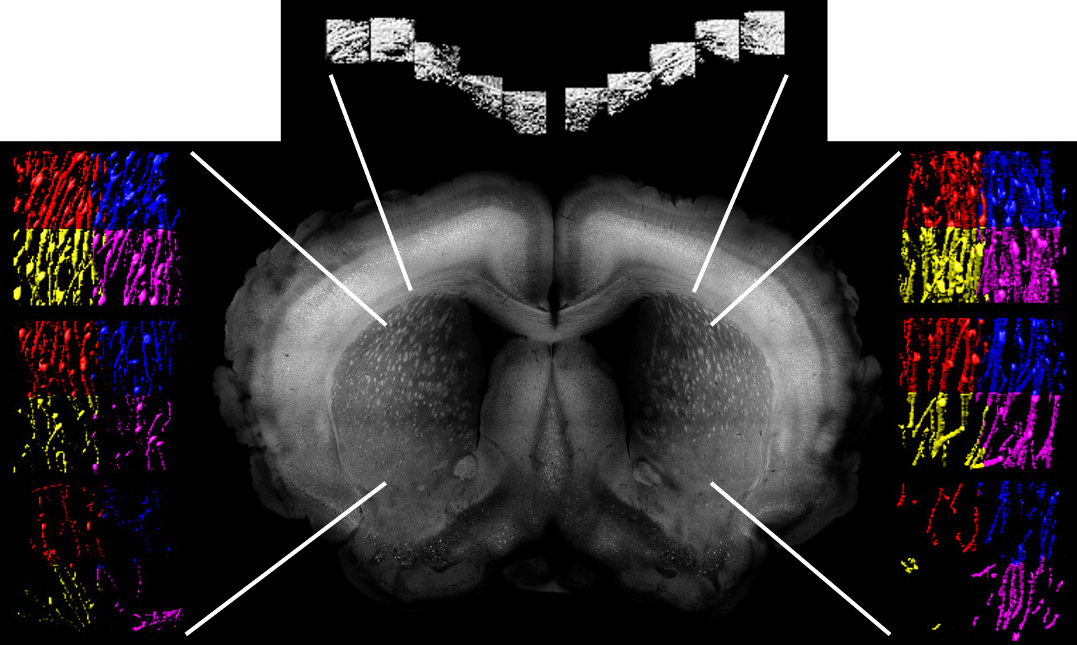
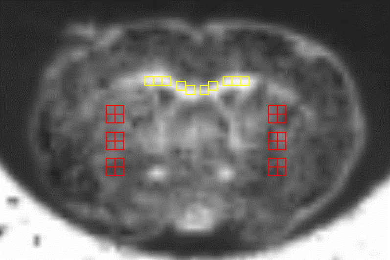
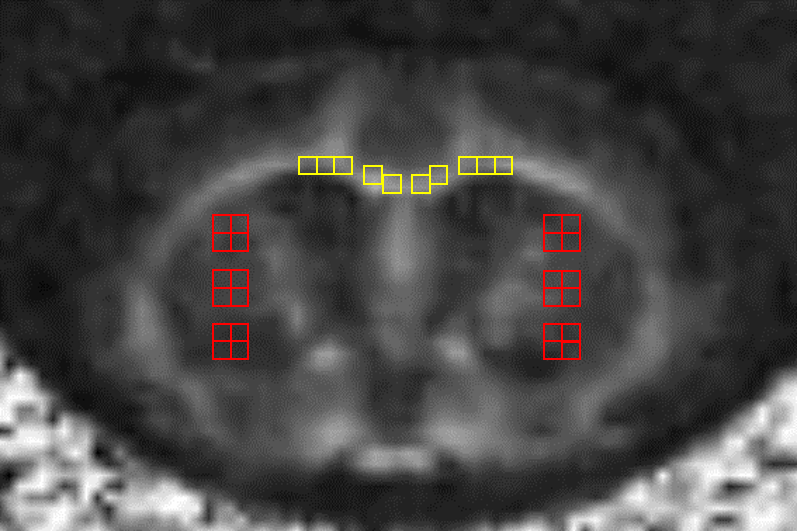
One thy-1 yellow fluorescent protein mouse was deeply anesthetized and perfusion fixation was performed. The brain was carefully dissected and whole brain MRI was acquired with a 7-T animal MRI system (Agilent Technologies Inc., Palo Alto, CA, USA). DKI and diffusion tensor imaging (DTI) data were obtained. After scanning MRI, brain sections were prepared with a thickness of 2mm and then cleared by using the clear, unobstructed brain imaging cocktails and computational analysis (CUBIC) method4. Confocal microscopy acquisition was performed with a Carl Zeiss LSM 780 two-photon microscope. Forty-eight regions of interest (ROIs) were set on the caudate nucleus and putamen and 16 ROIs were set on the corpus callosum in a confocal microscopy and a MR image (ROI size= 300×300×300µm). In each ROI, neurite density was calculated using Imaris Interactive Microscopy Image Analysis software (Bitplane, Zurich, Switzerland) with a threshold based surface reconstruction. The metrics of DKI and DTI such as mean, axial, and radial kurtosis (MK, AK, and RK), mean, axial, and radial diffusivity (MD, AD, and RD), and fractional anisotropy (FA) were computed by using dTV II and VOLUMEONE 1.81 (Image Computing and Analysis Laboratory, The University of Tokyo Hospital, Tokyo, Japan). The correlations between the diffusion metrics and neurite density were analyzed by Pearson correlation coefficients.Results
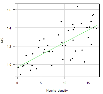
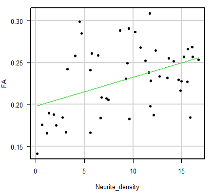
There was a strongly positive correlation between neurite density and MK (P<0.001, r=0.726), AK (P<0.001, r=0.621), and RK (P<0.001, r=0.737) in the caudate nucleus and putamen. The correlation between neurite density and FA in the caudate nucleus and putamen was moderate (P=0.003, r=0.420). Meanwhile, there was no significant correlation between neurite density and MK (P=0.971, r=0.010), and FA (P=0.172, r=0.359).Discussion
This study showed a strong positive correlation between MK and neurite density in the caudate nucleus and putamen. Especially, RK correlated more strongly with neurite density than AK, which was consistent with a known fact. The previous study showed that FA was very strongly correlated with neurite density in an area where nerve fiber orientations are unidirectionally aligned5. However, FA was not strongly correlated with neurite density in an area with crossing fibers such as caudate nucleus and putamen. Therefore, DKI was considered to be more sensitive to evaluate complex structures. In this study, the results of corpus callosum were not consistent. That was probably because the fluorescence in a confocal microscopy seems artificially low when close to the ventricle, and because neurite in the corpus callosum was very dense and hard to clarify in a microscopic image.Conclusion
DKI reflect neurite density in an area with crossing fibers, so that can evaluate more complex microstructures than DTI.Acknowledgements
This work was supported by the program for Brain Mapping by Integrated Neurotechnologies for Disease Studies (Brain/MINDS) from Japan Agency for Medical Research and development, AMED, and funded by ImPACT Program of Council for Science, Technology and Innovation (Cabinet Office, Government of Japan).References
1. Jensen JH, Helpern JA. MRI quantification of non-Gaussian water diffusion by kurtosis analysis. NMR Biomed 2010;23(7):698-710.
2. Hori M, Fukunaga I, Masutani Y, et al. Visualizing non-Gaussian diffusion: clinical application of q-space imaging and diffusional kurtosis imaging of the brain and spine. Magn Reson Med Sci 2012;11(4):221-233.
3. Kerever A, Kamagata K, Yokosawa S, et al. See-through Brains and Diffusion Tensor MRI Clarified Fiber Connections: A Preliminary Microstructural Study in a Mouse with Callosal Agenesis. Magn Reson Med Sci 2015;14(2):159-162.
4. Susaki EA, Tainaka K, Perrin D, et al. Whole-brain imaging with single-cell resolution using chemical cocktails and computational analysis. Cell 2014;157(3):726-739.
5. Kamagata K, Kerever A, Yokosawa S, et al. Quantitative Histological Validation of Diffusion Tensor MRI with Two-Photon Microscopy of Cleared Mouse Brain. Magn Reson Med Sci 2016.
Figures