3455
Cost effective 3D printed brain phantom for diffusion MRI1Dayananda Sagar Institutions, Bangalore, India
Synopsis
Diffusion weighted MRI is used to measure diffusion properties in the brain. In this paper, the method for the creation of an anatomically and mechanically realistic brain phantom from polyvinyl alcohol cryogel (PVA-C) and a 3D printable brain phantom using Poly Lactic Acid (PLA) PLA is proposed. PVA-C is material widely used in medical imaging phantoms because of its mechanical similarities to soft tissues. This brain phantom will allow testing and optimization of diffusion based MR methods.
Purpose
Diffusion Weighted Imaging (DWI) is a clinical tool to characterize isotropic and anisotropic water diffusion in the brain. Testing and optimization of MRI methods on human subjects is limited due to scanner availability. Thus, there is a need for “realistic” phantoms that will possess morphological features of the brain.1,2 The purpose of this study is to build a human brain phantom with anatomically realistic features and also investigate the characteristics of Poly Vinyl Alcohol (PVA) based diffusion MRI phantoms with different concentrations. The proposed phantom can be used to determine diffusion parameters similar to brain and can be utilized to optimize acquisition, reconstruction methods for diffusion MRI.Methods
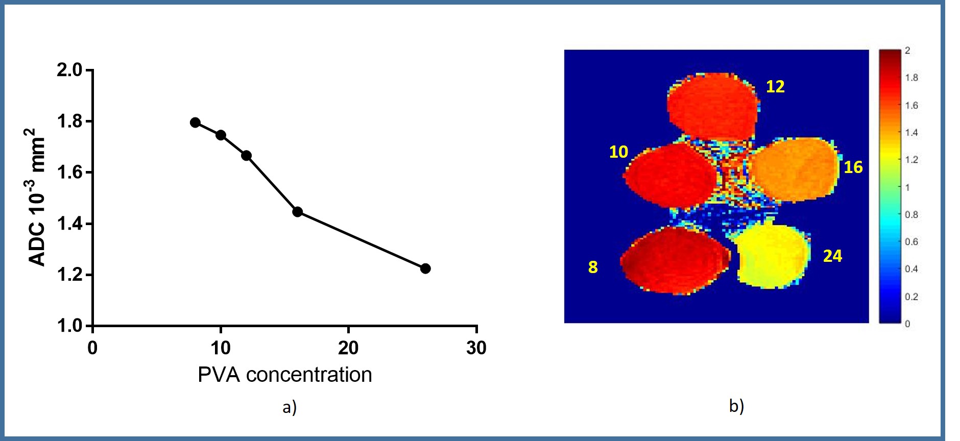
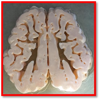
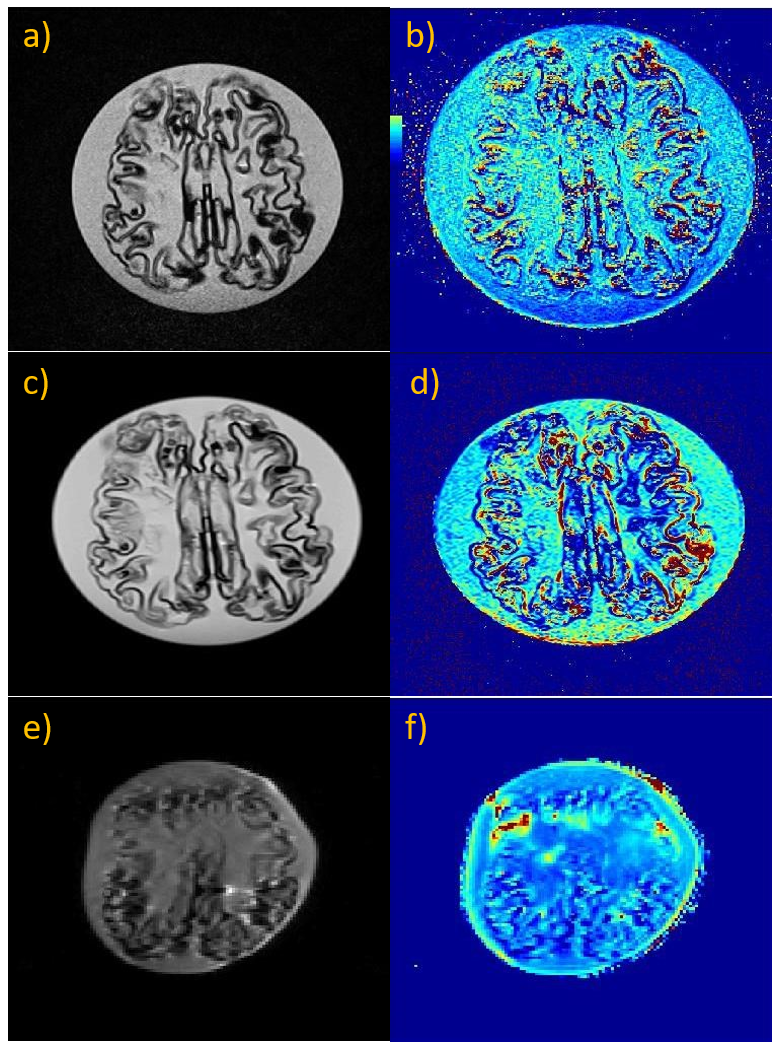
Phantom preparation: The human brain phantom consists of 2 parts, one is the exterior part of the brain made using Polylactic Acid (PLA) and the other is interior part of the brain mimicking the brain tissue using PVA. In our first experiment, PVA was prepared by dissolving fully hydrolysed PVA of 8, 10, 16, 20, 26 wt% by volume of water heating upto 1000° C. The mixture was heated till the solution was clear to get PVA cryogel. The PVA cryogel was cooled down gradually to room temperate with the lid closed so that the water content in cryogel is intact. The solutions were loaded into cylindrical plastic containers and cooled for 3 hours. Skeleton of the brain was printed with PLA material with the extruder temperature maintained at 210 °c and base plate temperature at 70° c from an open source 3D printable model. Brain phantom was placed in an oval shaped container filled with 12% wt by volume. MR Imaging: Series of MR images were acquired on a 1.5T Siemens scanner. DW imaging was performed using a single-shot spin echo imaging diffusion sequence; with 6 b-values varying from 0 to 2000 s/mm2 in 6 orthogonal directions and the corresponding trace was calculated. Images were acquired in coronal plane with a slice thickness of 4mm, an FOV of 200X 200 mm2, a matrix size of 128 × 128, and TR/TE: 3000/105ms. T1 maps coronal slices with variable flip angles (6 and 22) were obtained with the parameters [Slice thickness=3mm, TR/TE=20/4.4500ms]. T2 maps axial slices were obtained with the parameters [Slice thickness=3mm, TR/TE=2600/22ms, Averages=1].Results
The measured ADC values for concentrations 8,10,12,16 and 24wt % are 1.795, 1.746, 1.666, 1.446 and 1.224 X 10⁻³mm2/s respectively. ADC maps of varying concentration and single intensity plot can be seen in Fig. 1 with decreasing Signal Intensity (SI) curve. Fractional Anisotropy (FA) values for different diameters of 4, 5.5, 6.5,7 and 7.5cms are 0.114, 0.119, 0.169, 0.062and 0.0454. Fig 2 shows axial slice of a 3D printed brain. Fig 3(a-d) shows T1 and T2 weighted maps respectively. It can be observed that T1 and T2 weighted images shows a brain structure, which resembles the human brain. Fig 3(e-f) shows DW image and ADC map, it can be observed the noisy image which is attributed to gradients. Overlapping and geometric distortion can be seen on ADC and FA maps as part of EPI and phase over sampling.Discussion and conclusion
PVA’s diffusion values are in range of human brain (gray matter: 0.7- 0.85, white matter: 0.67-0.8, CSF- 3-4mm2/s). Decrease in concentration of PVA gives higher ADC values, which can be used to prepare different phantoms mimicking specific tissue properties. This is attributed to with increase in concentration of PVA, molecules arrange more closely. In this work, it has also been illustrated that PVA’s anisotropy value decreases with increase in diameter, this property plays a significant role in determining anisotropy nature and modeling it to mimic white matter in human brain. PVA is not expensive and can be used as a material for brain phantom to study diffusion properties.Acknowledgements
This work was supported by Department of Electronics and Information Technology (DEITY/1(15)-2014 ME& HI)References
[1] Surry, K. J. M., Austin, H. J. B., Fenster, A., & Peters, T. M. (2004). Poly(vinyl alcohol) cryogel phantoms for use in ultrasound and MR imaging. Physics in Medicine and Biology, 49(24), 5529–5546. Evaluation Studies, Journal Article, Research Support, Non-U.S. Gov’t, Validation Studies.
[2] Chen, S. J.-S., Hellier, P., Marchal, M., Gauvrit, J.-Y., Carpentier, R., Morandi, X., & Collins, D. L. (2012). An anthropomorphic polyvinyl alcohol brain phantom based on Colin27 for use in multimodal imaging. Medical Physics, 39(1), 554–561. Journal Article, Research Support, Non-U.S. Gov’t. http://doi.org/10.1118/1.3673069
[3] Web: www.thingiverse.com/: Source for the STL file
Figures