3446
Water-fat MRI demonstrates seasonal proliferation of brown adipose tissue near the eyes of juvenile hibernators: An additive effect of cold exposure1Biology, University of Western Ontario, London, ON, Canada, 2Medical Biophysics, University of Western Ontario, London, ON, Canada
Synopsis
Hibernating mammals use brown adipose tissue (BAT) as a primary source of heat production. Volumes of both white adipose tissue (WAT) and BAT increase in the autumn even when temperatures are warm. Between Aug 19th and Oct 13th we used water-fat MRI to measure the dynamics of BAT and white adipose tissue as ground squirrels prepared for hibernation under either cold or thermoneutral temperatures. We found that the volume of a tissue that resembles BAT around the eye, increased significantly in cold exposed animals to warm exposed in October, as the animals are preparing for winter.
Target Audience
This abstract is targeted to those interested in MRI of brown adipose tissue.Introduction
During the winter, hibernating mammals cycle between periods with very low body temperature (~5°C) that last for several days, and brief arousal periods with normothermic body temperature near 37°C. During these spontaneous arousals, brown adipose tissue (BAT) is the primary source of heat production. In non-hibernating mammals, proliferation of BAT requires extensive acclimation to cold environmental temperatures. In contrast, expression of BAT-specific genes in hibernators follows an endogenous rhythm, increasing in autumn even when animals are held at constant, warm temperatures1. Recently we demonstrated that BAT in the thorax of ground squirrels increases in volume without cold exposure in anticipation of hibernation 2. We also described a tissue near the eyes with a fat fraction that resembles closely thoracic BAT. The brain temperature of hibernating ground squirrels is maintained about 3.7°C above the core body temperature3. We hypothesize that volumes of this “eye BAT” would increase as the hibernation season approached, and that cold exposure would accelerate this proliferation.Purpose
To assess the effect of cold versus warm exposure on the volume of BAT-like tissue in squirrels surrounding the eyes in anticipation for hibernation using water-fat MRI.Methods
Two groups of four male 13-lined ground squirrels (Ictidomys tridecemlineatus) were housed at constant photoperiod (12h light, 12h dark) but at different temperatures: 25°C and 5°C. The squirrels were scanned using a 3T MRI scanner (Discovery MR750, GE Healthcare, Waukesha, WI, USA) and a 32 channel cardiac coil under a protocol approved by the institution’s Animal Care Subcommittee. Animals were anaesthetized using isoflurane and 100% oxygen and scanned in August and again 4 and 7 weeks after the initial scan. T1-and T2-weighted images were acquired (TR/TE/flip angle/number of averages = 4.3ms/2.0ms/15°/4 and 2000ms/162ms/90°/2, respectively, with voxel dimensions = 0.9mm isotropic for both acquisitions). IDEAL water-fat images (reconstructed with correction for T2* decay and fat spectral complexity) were also collected with TR/∆TE/# echoes/flip angle = 7.96ms/0.856ms/6/4° and voxel dimensions = 0.9mm isotropic. The T1-weighted images were used to manually segment total squirrel volume. BAT has a lower fat fraction than White Adipose Tissue4, so fat fraction images generated from the water-fat images were used for semi-automatic segmentation of BAT surrounding the eyes. All segmentation was performed by (AM) using the “2D growing region” tool of OsiriX v.5.8.2 (Pixmeo, Geneva, Switzerland). Repeated measures two-way ANOVA was used to test the null hypothesis that BAT volume did not change over the 7 weeks, and that cold exposure did not affect BAT volume.Results
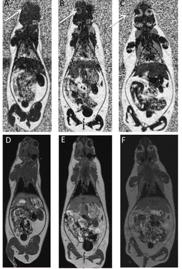
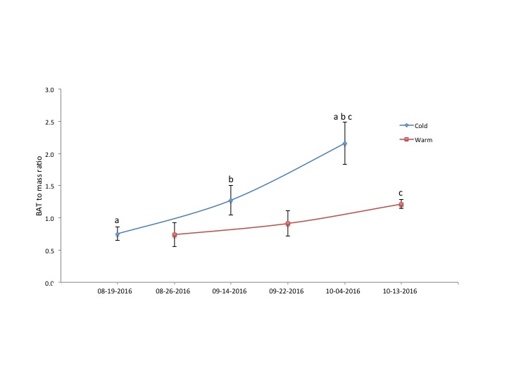
Figure 1a shows a representative fat fraction image at three time points with the BAT locations indicated. Figure 1b shows corresponding T1 weighted images. Figure 2 demonstrates a significant increase in eye BAT volume when acclimated to cold. On Oct 13th there is significant increase in volume of eye BAT between warm and cold squirrels (P=0.006). The mean eye BAT volume with cold acclimation increased 2.8 fold, and warm 1.6 fold during the 7 weeks between imaging sessions.Discussion
In most mammals cold exposure is necessary for BAT proliferation, but our data show that BAT volume increases in hibernators held at thermoneutral temperatures. Cold exposure has an additive effect on BAT proliferation surrounding the eyes. This is the first report of a tissue resembling BAT around the eyes. It is remotely possible that this tissue is actually the Harderian gland that, in mice is comprised of 35% lipid (w/w), which is within the fat fraction values we used to identify BAT (i.e. 25%-70%). The chemical lipid extraction used to assess Harderian gland lipid composition5 detects virtually all lipids, including lipids found in the membranes. Water-fat MRI, on the other hand, only detects lipids that are 'mobile', especially triglycerides, but not lipids found within membranes including myelin6. We believe, therefore, that the MRI-detectable fat fractions from this gland would be lower than that detected by chemical extraction. In fact, we expect the fat fraction of this gland to fall below the lower 25% threshold to be detected by our water-fat MRI technique. Finally, it is unlikely that there would be a 2.8-fold increase volume of the Harderian gland prior to hibernation.Conclusion
This water-fat MRI study shows, first time that a BAT (based on fat fraction) was discovered behind the eyes and that cold exposure can have an additive effect in proliferation of BAT in hibernators.Acknowledgements
The authors acknowledge support from the Natural Sciences and Engineering Research Council.References
1. Hindle AG and Martin SL. Intrinsic circannual regulation of brown adipose tissue form and function in tune with hibernation. Am J Physiol Endocrinol Metab. 2014 306(3):E284
2. Amanda MacCannell, Kevin Sinclair, Lanette Friesen-Waldner, et al. Water-Fat MRI Detects Increased Brown Adipose Tissue Volume In Anticipation of Hibernation in Squirrels [Abstract]. Proceedings of the 24th Annual meeting of ISMRM; 2016. Abstract nr 3930.
3. Barger JL, Barnes BM, Boyer BB. Regulation of UCP1 and UCP3 in arctic ground squirrels and relation with mitochondrial proton leak. J Appl Physiol. 2006 101(1): 339–347.
4. Hu HH, et al. Identification of brown adipose tissue in mice with fat-water IDEAL-MRI. J Magn Reson Imaging
5. Watanabe M. An autoradiographic, biochemical, and morphological study of the Harderian gland of the mouse. J Morphol. 1980 163:349–365.
6. Hines CDG, Yu H, Shimakawa A, et al. Quantification of hepatic steatosis with 3-T MR imaging: validation in ob/ob mice. 2010 Radiology 254:119–128.
Figures