3445
Validity of estimating subcutaneous and visceral fat volume from single MRI slice in older adults with sarcopenia and sarcopenic obesity1Institute of Geriatrics and Active Ageing, Tan Tock Seng Hospital, Singapore, Singapore, 2Geriatric Medicine, Tan Tock Seng Hospital, Singapore, Singapore, 3Diagnostic Radiology, Tan Tock Seng Hospital, Singapore, Singapore
Synopsis
The demand for measurements of fat quantities is driven by the rising prevalence of sarcopenia and sarcopenic obesity (SO). In order to reduce the time and the cost of image processing, several studies have estimated the subcutaneous fat (SF) and visceral fat (VF) volume from a single slice. However, the population of studies may not have necessarily included patients with either sarcopenia or SO. This study aims to determine the correlation between the cross-sectional areas in a single slice at different vertebra levels and the volumes of SF and VF in the abdomen for sarcopenic and SO populations using MRI.
Introduction
Magnetic resonance imaging (MRI) is a promising imaging modality to quantify muscle and fat volume, especially for sarcopenia and sarcopenic obese (SO) subjects, given its ionizing radiation-free nature is suitable for scanning older adults. Subcutaneous fat (SF) and visceral fat (VF) volume can be measured by segmenting muscles and fat tissues from a series of MRI images covering a region of interest (ROI). However, this method is tedious and time-consuming as multiple structures in tens of slices need to be segmented to cover the entire ROI. In order to reduce the time and the cost of image processing, several studies have proposed to estimate SF and VF from a single slice1-4. Various vertebra locations have been reported that correlated best with the SF and VF volume of the entire abdomen. However, the population of studies may not have necessarily included patients with either sarcopenia or SO. Furthermore, the majority of published data has been based on CT images. The objectives of this study was to determine if there was correlation between the cross-sectional areas (CSAs) in a single slice at different vertebra levels and the volumes of SF and VF in the abdomen for sarcopenic and SO populations using MRI.Methods and Materials
MRI scans of the thigh of 190 healthy community dwelling older adults were acquired and analyzed as a part of a larger longitudinal study. Dixon GRE sequences were acquired and SF and VF were automatically segmented in the abdomen region between L2-L4 lumbar vertebra using a fully automated graph theoretic segmentation algorithm5. Volumes of SF and VF were calculated for the entire L2-L4 lumbar vertebra volume and CSAs were assessed for slices at L1/L2, L2/L3, L3/L4, and L4/L5 vertebra levels in each subject.Results
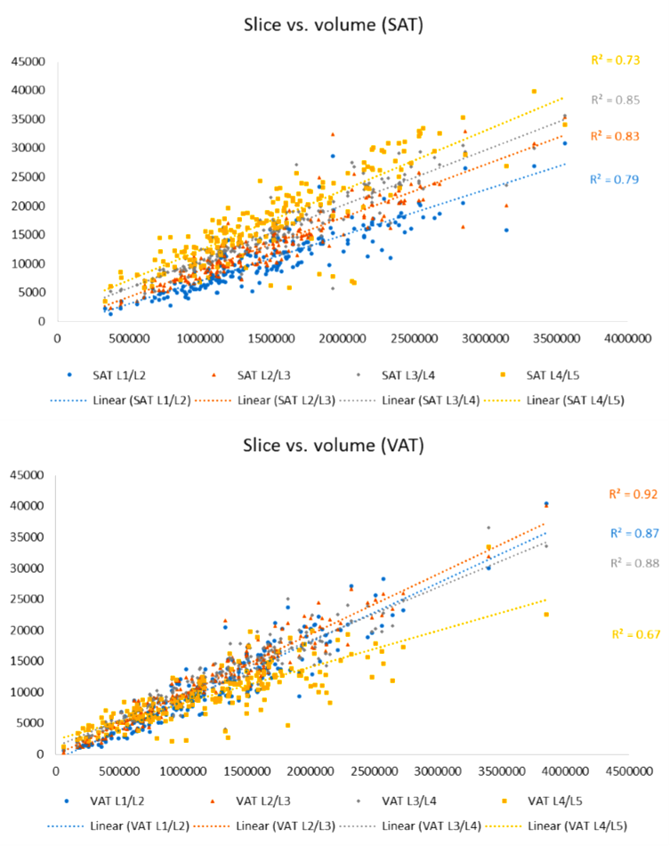
Fig. 1 shows the linear regressions between abdomen volume and CSAs at different levels of the vertebra for SF and VF. For SF, the correlation of determination (R2) for linear regressions between the CSAs at L2/L3 and L3/L4 levels were high, 0.83 for L2/L3 and 0.85 for L3/L4. For VF, the highest R2 was at L2/L3 level (0.92).Conclusion
A single CSA at L2/L3 vertebra level yields good estimation of SF and VF volume in abdomen for older adults with sarcopenia and SO. This has potential to greatly reduce scan time and imaging costs, for future implementation in routine clinical practice.Acknowledgements
No acknowledgement found.References
1. Shen W, Punyanitya M, Chen J et al (2007) Visceral adi- pose tissue: relationships between single slice areas at different locations and obesity-related health. Int J Obes (Lond) 31:763–769
2. Irlbeck T, Jm Massaro, Bamberg F, O’Donnel CJ, Hoffman U, ?Fox CS (2010) Association between single-slice measurements of visceral and abdominal subcutaneous adipose tissue with vol- umetric measurements: the Framingham Heart Study. Int J Obes (Lond) 34:781–787 ?
3. Kuk JL, Church TS, Blair SN, Ross R (2006) Does measure- ment site for visceral and abdominal subcutaneous adipose tis- sue alter associations with the metabolic syndrome? Diabetes Care 29:679–684 ?
4. Kuk JL, Church TS, Blair SN, Ross R (2010) Measurement site and the association between visceral and abdominal subcutane- ous adipose tissue with metabolic risk in women. Obesity (Sil- ver Spring) 18:1336–1340
5. Parimal SA, Zagorodnov V. Segmentation of magnetic resonance images of brain and abdomen. Masters Thesis, Nanyang Techno- logical University, Singapore; 2010.
Figures