3425
Comparison between IVIM combined with fuzzy clustering algorithm and IVIM combined with Bayesian method in the thyroid cancerKaining Shi1, Fengmao Chiu2, Yunlong Yue3, Lee Jiang4, Lili Zuo3, and Yanfang Jin3
1Philips Healthcare (China), Beijing, People's Republic of China, 2Philips Healthcare, Taipei, Taiwan, 3Department of MR, Beijing Shijitan hospital of capital medical university, Beijing, People's Republic of China, 4Philips Healthcare (China), Suzhou, People's Republic of China
Synopsis
To improve the stability of the nonlinear curve fitting of IVIM, the Fussy clustering technique (FCM) and data driven Bayesian approach are combined with IVIM in the imaging of thyroid tumors. Both FCM and Bayesian approach can improve the homogeneity of IVIM parameters. With limited sample size, FCM has similar diagnosis efficiency with conventional method using less calculation time, while Bayesian approach doesn’t increase the diagnosis efficiency.
Introduction
Intravoxel Incoherent Motion (IVIM) imaging, which mainly is the bi-exponential analysis of the multiple b values DWI, has been widely applied in many applications. One drawback of IVIM is its poor stability, especially for perfusion related parameters f and D*. To address this problem, the Fussy clustering technique (FCM) has been combined with the nonlinear curve fitting of IVIM1-2. Data driven Bayesian approach also has the ability to improve the robustness of IVIM by using a shrinkage prior model3. However, none of above approaches have been evaluated in the diagnosis of diseases.Purpose
To compare the efficiency of FCM-IVIM with IVIM using Bayesian approach in the diagnosis of thyroid cancer.Method
10 patients with thyroid tumors were scanned by a 3T whole body scanner ( Ingenia, Philips Medical System, Best, The Netherlands) using a 8-ch carotid coil. 5 malignant and 6 benign tumors were confirmed through the biopsy. A single-shot spin echo DWI using 2D RF pulse was scanned with following parameters: TE/TR 69/1400ms, FOV 160x47mm, acquisition matrix 108x101, 10 slices with the thickness of 5mm and 1mm gap, NSA 4. 8 b values (0, 20, 50, 100, 200, 400, 600, 990) were used. The non-linear fitting of the bi-exponential model was performed on home-made software. Mask for calculation was restricted within the thyroid. Total cluster number used in the FCM is 10 multiplied by slice number. 3D ROI was manually drawn on multiple slices to cover the whole tumor. Average values of IVIM derived parameters ( D, D* and f) of the whole tumor were used for each approach. Friedman test was employed to compare IVIM parameters of FCM and Bayesian with those calculated pixel by pixel (P<0.05). Two samples T test was performed to evaluate the efficiency of each approach in differentiating benign from malignant tumors (P<0.05).Result
Both FCM and Bayesian methods can improve the homogeneity of values in the lesions (Fig 1&2). D and D* derived from Bayesian methods is different with FCM or pixel by pixel method ( F=33.2143, P <0.00001; F= 18.8095, P=0.00003), f from any method is different with others (F=40.4167, P <0.00001) (Fig.3). Both D derived from pixel by pixel and from FCM methods have significant difference between benign group and malignant group (P = 0.0015, P = 0.0105), while f and D* don’t( P = 0.4879 and P = 0.5954 for f, P = 0.3164 and P = 0.7811 for D*). None of three parameters from Bayesian method exhibits significant difference between two groups (P = 0.0554 for D, P = 0.1970 for f, P = 0.6549 for D*) (Fig.4).Discussion
Bayesian approach can improve the homogeneity of IVIM parameters. However, it changed the IVIM parameters too much from the conventional method, especially even the trend of D* between benign and malignant tumors. FCM has similar results with conventional method, with minimal cost of time. Though the lack of ‘gold standard’ of IVIM makes numerical parameter comparison meaningless, the result of this study doesn’t exhibit any improvement of the diagnosis efficiency of two purposed method. One reason is that the 3D ROI covered whole tumor may increase the stability of conventional approach. Another reason may be the limited sample size. More data needs to be involved in the further work.Conclusion
Both FCM and Bayesian approach can improve the homogeneity of IVIM parameters. With limited sample size, FCM has similar diagnosis efficiency with conventional method using less calculation time, while Bayesian approach doesn’t increase the diagnosis efficiency.Acknowledgements
No acknowledgement found.References
1. K Shi, et al. ISMRM (2015) 2905. 2. K Shi, et al. ISMRM (2016) 3467. 3. MR Orton, et al. MRM 2014;71(1):411-20Figures
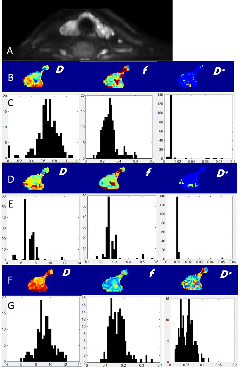
A 24 years old female patient with malignant tumor on
the right side of thyroid. (A) DWI b=990; (B) D, f and D* maps derived from
conventional method, and corresponding histograms of the whole tumor (C); (D)
D, f and D* maps derived from FCM method, and corresponding histograms of the
whole tumor (E); (F) D, f and D* maps derived from Bayesian method, and corresponding
histograms of the whole tumor (G);
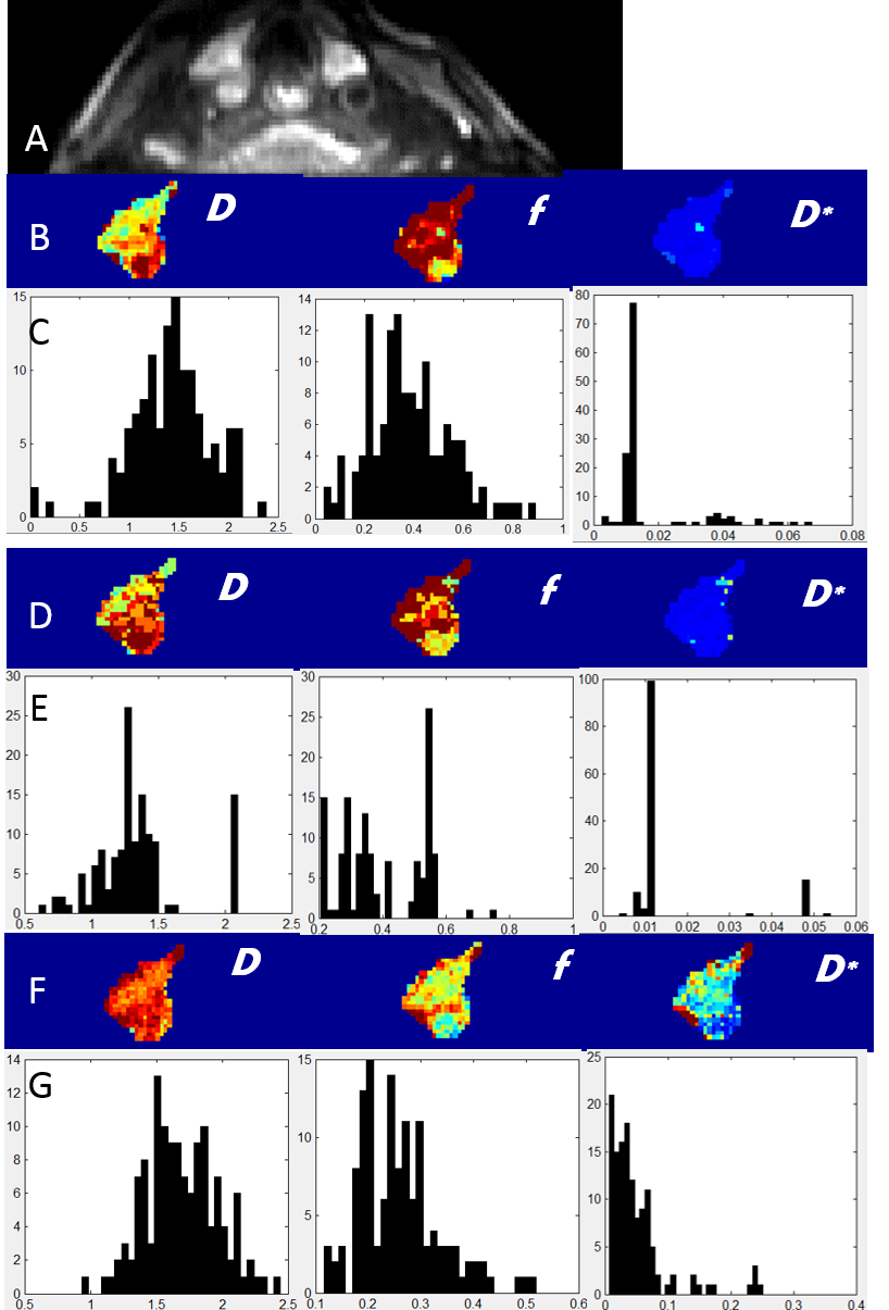
A
62 years old female patient with benign tumor on the right side of thyroid. (A)
DWI b=990; (B) D, f and D* maps derived from conventional method, and corresponding
histograms of the whole tumor (C); (D) D, f and D* maps derived from FCM
method, and corresponding histograms of the whole tumor (E); (F) D, f and D*
maps derived from Bayesian method, and corresponding histograms of the whole
tumor (G);
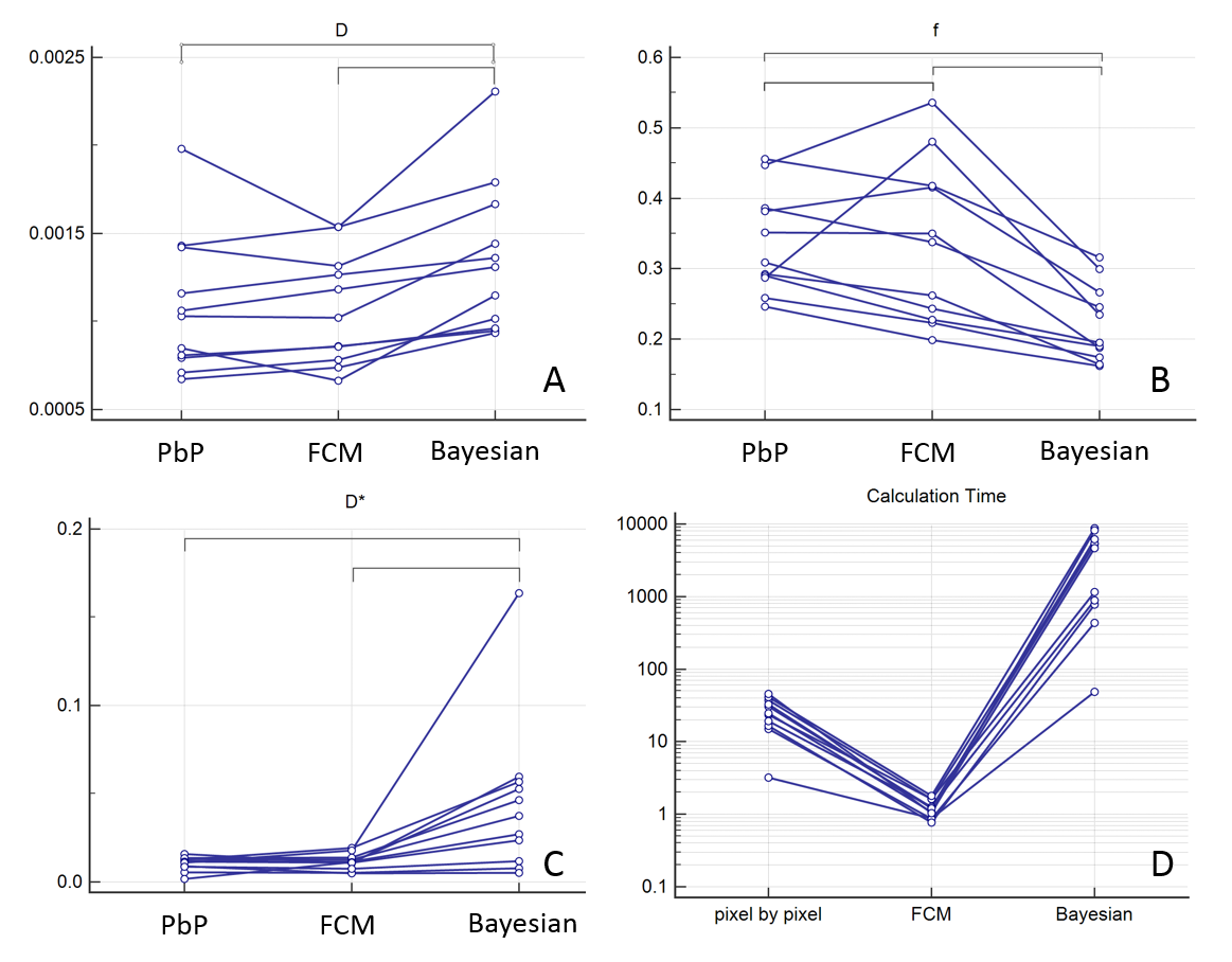
D(A),f(B),D*(C)
values and total calculation time (D) of all 11 lesions with three methods. Statistical
difference of values from different methods is indicated with connectors. Note
that time difference is huge so the logarithmic display of the vertical axis is
used.
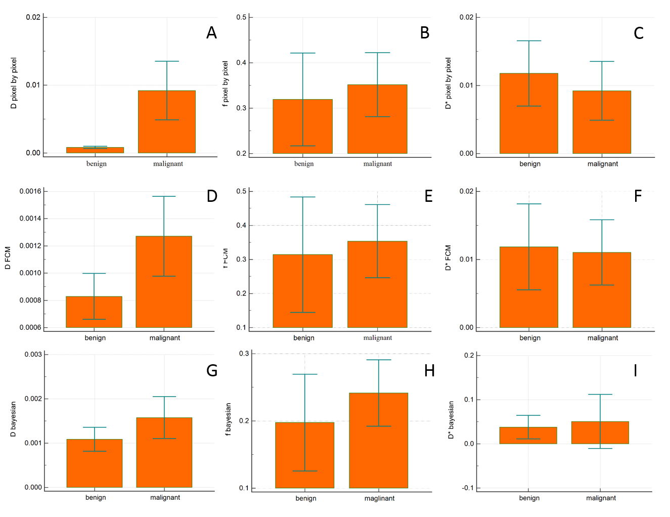
IVIM
parameters of the benign and malignant group. Only D of conventional (A) and
FCM(D) approach has statistical significant.