3421
Simultaneous multislice diffusion weighted imaging in whole-body PET/MRI for accelerated multiparametric staging of oncologic patients.1Department of Diagnostic and Interventional Radiology, Eberhard-Karls-University Tuebingen, Germany, Tuebingen, Germany, 2Section on Experimental Radiology, Eberhard-Karls-University Tuebingen, Germany, Tuebingen, Germany
Synopsis
Simultaneous multislice diffusion weighted imaging (SMS-DWI) is a promising technique to shorten scan time in MRI. Aim of our study was to compare the diagnostic performance of SMS-DWI to conventional DWI for multiparametric whole-body examinations of oncologic patients in PET/MRI. We performed an evaluation in three steps: First in a phantom study, second in a volunteer study and third in a patient study with 20 oncologic patients. We found that SMS-DWI led to a significant reduction of scan time and, although suffering from slightly impaired image quality, provided reliable ADC values and lesion conspicuity of PET positive lesions.
Introduction
Positron emission tomography/magnetic resonance imaging (PET/MRI) is a hybrid imaging modality which is able to acquire PET and MRI simultaneously and can therefore provide a multiparametric tumor assessment. In this connection, diffusion weighted imaging (DWI) plays an essential role in MRI as it is able to sensitively detect and quantify changes of tissue diffusivity [1]. Multiparametric whole-body examinations in PET/MRI can take 60-90 minutes which not only limits its acceptance in patients but hampers also its use in daily clinical routine [2]. Recent technical developments enabled simultaneous multiband radiofrequency excitation and acquisition of multiple slices [3-5] which reduces the scan time by the number of simultaneously excited slices. The potential of simultaneous-multi-slice (SMS)-DWI to reduce examination time in whole-body imaging has been shown in previous studies [6]. Aim of our study was to compare the diagnostic performance of SMS-DWI to conventional DWI (C-DWI) for multiparametric whole-body examinations of oncologic patients in PET/MRI.Material and Methods
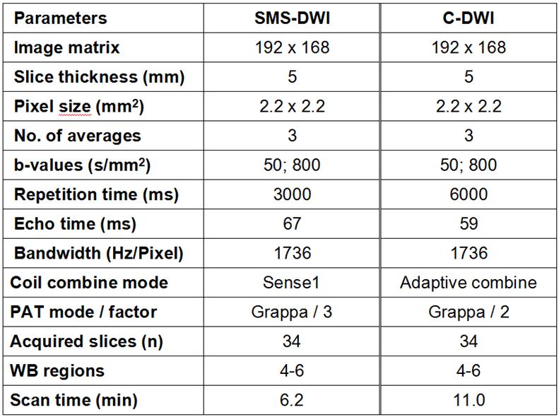
The study was approved by the local ethic committee. Examinations were performed on a 3 Tesla PET/MRI scanner (Biograph mMR, Siemens Healthineers, Erlangen, Germany). Sequence parameters for SMS-DWI and C-DWI are given in table 1. We performed a comparison of SMS-DWI and C-DWI in three steps: First in a phantom study by acquiring the signal-to-noise ratio (SNR) and the mean values of the apparent diffusion coefficient (ADC) in solutions of different diffusivities (0-40% glucose concentration). Second in a volunteer study (n=10, 29±9.3 years, 5 female) by I) assessing the ADC in different body regions (white matter, liquor, right and left liver lobe, spleen, kidney and psoas muscle) and II) evaluating the overall image quality using a 5-point Likert-scale (1=poor, 5=excellent) in different body regions (head, neck, thorax, upper abdomen, pelvis) by two independent radiologists in b=800 images and ADC images. And finally in an evaluation of 20 oncologic patients (62.3±10.3 years, 7 female). All patients received a whole-body examination with a PET scan, both DWIs and morphologic sequences (e.g. post-contrast T1 VIBE or T2 HASTE). We evaluated I) the overall image quality in the same manner as in the volunteer study, additionally with categorizing the observed artifacts (distortion, fat sat, ghosting, others) as well as assessing the overall image noise and image sharpness; II) the ADC of PET positive metastatic lesions >1cm in diameter and III) the conspicuity of PET positive lesions (max. 5 per organ) in the b=800 image (1=not visible, 5=excellent). Evaluations were performed with the software MMOncology (syngo.via, Siemens Healthineers, Erlangen, Germany). Statistical analysis were performed with a pairwise Dunn-Bonferroni post hoc tests in the Wilcoxon signed rank test using the software SPSS (v. 23.0; IBM, Chicago, IL). p-values < 0.05 were considered significant.Results
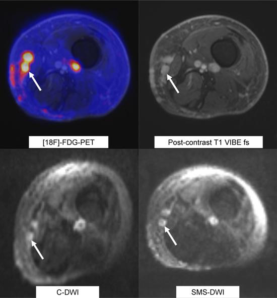
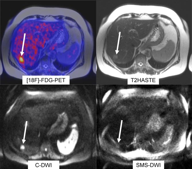
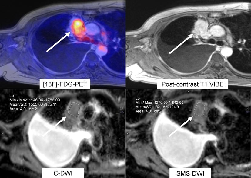
Analysis of ADC values of different concentration of glucose solution revealed only marginal differences of about 0.3-1.3% between SMS-DWI and C-DWI sequences. The evaluation of DW images showed about of 47% lower SNR in SMS-DWI compared to C-DWI. Using SMS-DWI for whole-body examinations, the scan time could be reduced by 44% compared to C-DWI (table 1). In the volunteer and patient studies the subjective image quality did not differ significantly between SMS-DWI and C-DWI except for the thoracic region and the upper abdominal regions where the image quality of SMS-DWI in b=800 images as well as in ADC images were rated inferior (3.0 and 3.8, p=0.02). ADC values of SMS-DWI were significantly lower in the right and left liver lobe (1.0 and 0.9; 1.4 and 1.1) and the psoas muscle (1.4 and 1.3). DWI images with b=800 and ADC images of SMS-DWI provided a higher image sharpness (ADC: 3.8 and 3.2, p<0.01, figure 1) while image noise was also rated higher. N=118 lesions were evaluated for lesion conspicuity and no significant difference could be found although a trend towards lower conspicuity of lesions located in the thoracic and upper abdominal region was seen in SMS-DWI (figure 2). ADC values of PET positive lesions >1cm did not differ significantly (n=28, p=0.86, figure 3). More artifacts were seen in the SMS-DWI in all categories except for ghosting artifacts in b=800 images.Conclusion
SMS-DWI
provided higher image sharpness and reliable conspicuity and ADC values of PET
positive tumorous lesions. Therefore, although suffering from lower SNR and
impaired image quality especially in the thoracoabdominal region, SMS-DWI has
the potential to distinctly reduce scan time for oncologic patients in multiparametric
whole-body PET/MRI examinations.Acknowledgements
No acknowledgement found.References
1. Padhani, A.R. and D.M. Koh, Diffusion MR imaging for monitoring of treatment response. Magn Reson Imaging Clin N Am, 2011. 19(1): p. 181-209.
2. Guckel, B., et al., Patient comfort during positron emission tomography/magnetic resonance and positron emission tomography/computed tomography examinations: subjective assessments with visual analog scales. Invest Radiol, 2015. 50(10): p. 726-32.
3. Feinberg, D.A., et al., Multiplexed echo planar imaging for sub-second whole brain FMRI and fast diffusion imaging. PLoS One, 2010. 5(12): p. e15710.
4. Breuer, F.A., et al., Controlled aliasing in parallel imaging results in higher acceleration (CAIPIRINHA) for multi-slice imaging. Magn Reson Med, 2005. 53(3): p. 684-91.
5. Moeller, S., et al., Multiband multislice GE-EPI at 7 tesla, with 16-fold acceleration using partial parallel imaging with application to high spatial and temporal whole-brain fMRI. Magn Reson Med, 2010. 63(5): p. 1144-53.
6. Kenkel, D., et al., Whole-Body Diffusion Imaging Applying Simultaneous Multi-Slice Excitation. Rofo, 2016. 188(4): p. 381-8.
Figures