3419
The ADC characteristics of the bone marrow within different anatomical parts: a DWI MR based study1Department of Radiology, Tianjin first center hospital, Tianjin, People's Republic of China, 2Department of Radiology
Synopsis
The diffusion weighted imaging has been widely used in clinical disease diagnosis, and the ADC value plays a key role in this progress. At present, the ADC values of normal organs and tissues have been reported, but few studies have been done on ADC values of normal bone marrow within different anatomical parts. In this study, the ADC values within different anatomical parts of bone marrow were measured and analyzed by using WB-DWI (whole body diffusion weighted imaging). And we furtherly evaluated the relationship of these ADC values with age and gender.
Purpose
The diffusion weighted imaging has been widely used in clinical disease diagnosis, and the ADC value plays a key role in this progress. At present, the ADC values of normal organs and tissues have been reported [1-3], but few studies have been done on ADC values of normal bone marrow within different anatomical parts. In this study, the ADC values within different anatomical parts of bone marrow were measured and analyzed by using WB-DWI (whole body diffusion weighted imaging). And we furtherly evaluated the relationship of these ADC values with age and gender.Methods
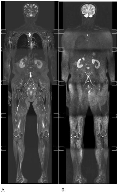
Thirty-five normal volunteers (17 male and 18 female, aged 21-82 years) were included in this study and scanned with WB-DWIBS protocol based on a 3T MR scanner (Ingenia ,Philips Healthcare, Best, the Netherlands). The DWIBS (2 b-values of 0 and 800 s/mm2) and STIR T2-TSE (Fig.1) were performed with a multi-station acquisition for whole body coverage with head and neck coil, two anterior torso coils and built-in posterior coil. The total scan time was within 50 minutes for each subject. The ADC values within ROIs located at the bone marrow of the thoracic spine, lumbar spine, sternum, clavicle, iliac wing, femoral head, femoral neck, proximal femur, tibia and fibula were measured respectively. The ADC values within different ROIs were compared by the one-way ANOVA. Besides two sample Student’s t-test were performed for the each ROI between male and female groups to find whether there are some differences between different sexes. Meanwhile, correlation analysis was carried out to investigate the relationships between ADC values of different ROIs with the age and gender.Result
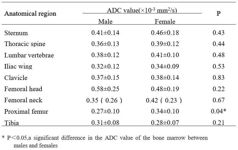
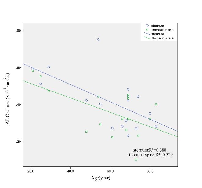
The bone marrow ADC values of thoracic spine, lumbar spine, sternum, clavicle, iliac wing, femoral head, femoral neck, proximal femur, tibia and fibula were summarized in Table 2. There was a significant difference in the ADC value of the bone marrow between males and females in the proximal femur (p=0.04). The ADC of bone marrow was significantly higher in female than in male group. No significant difference was found in other ROIs. Correlation analysis results showed that the ADC value of thorax, sternum in male groups have a negatively correlation with age(r=0.62 and 0.60 and p=0.01 and 0.02 respectively)in Table 3.Discussion
The ADC value of bone marrow reflects the diffusion characteristics of different anatomical parts. And it may vary with ages and genders. The results can provide the basis for the evaluation of the bone marrow characteristics with different anatomical parts and the diagnosis of bone marrow lesions.Conclusion
Bone marrow ADC values of proximal femur have a significant difference between males and females. There are some correlations between the ADC value of thorax spine and sternum with age respectively. The results might provide a basis for further study of multiple myeloma, bone metastases and other systemic bone marrow lesions by using DWI imaging.Acknowledgements
No acknowledgement found.References
1. Li Q, Pan SN, Yin YM, et al. Normal cranial bone marrow MR imaging pattern with age-related ADC value distribution. Eur J Radiol 2011;80:471–477.
2. Herrmann J, Krstin N, Schoennagel BP, et al. Agerelated distribution of vertebral bone-marrow diffusivity. Eur J Radiol 2012;81:4046–4049.
3. Lavdas I., et al., Apparent Diffusion Coefficient of Normal Abdominal Organs and Bone Marrow From Whole-Body DWI at 1.5 T: The Effect of Sex and Age. AJR Am J Roentgenol, 2015. 205(2): p. 242-50.
Figures