3403
Diffusion-Weighted Split-Echo RARE Imaging Free Of Geometric Distortion for Renal MRI at Ultrahigh Fields1Berlin Ultrahigh Field Facility (B.U.F.F.), Max Delbrueck Center for Molecular Medicine, Berlin, Germany, 2Institut für Vegetative Physiologie, Charité - Universitätsmedizin Berlin, Berlin, Germany, 3Physikalisch-Tecnische Bundesanstalt (PTB), Berlin, Germany, 4Experimental and Clinical Research Center (ECRC), a joint cooperation between the Charité Medical Faculty and the Max Delbrueck Center for Molecular Medicine in the Helmholtz Association, Berlin, Germany
Synopsis
T2* mapping does not fully represent renal tissue oxygenation. Diffusion-weighted imaging (DWI) can provide information about confounding factors, which can be used to correct T2*. The most widely used DWI technique SE-EPI is sensitive to magnetic field inhomogeneities and hence prone to geometric distortions. In this work we propose a diffusion-weighted Rapid Acquisition Refocusing Enhancement (RARE) variant for DWI of the rat kidney free of geometric distortions. Phantom experiments validated the diffusion weighting implementation in the common RARE sequence. Ex-vivo and in-vivo experiments using diffusion-weighted RARE showed no geometric distortions at 9.4 Tesla.
Introduction
The imbalance between oxygen demand and oxygen supply is believed to be a cause of several kidney diseases. Blood oxygenation sensitized MRI (T2*mapping) can non-invasively provide information about changes in renal oxygenation. Yet, previous experiments combining non-invasive MRI and invasive physiological measurements of the kidney obtained under different (patho)physiological conditions showed that T2* does not accurately represent renal tissue oxygenation1,2. Confounding factors such as tubular volume fraction should be taken in account for the interpretation of renal T2* mapping and for a reliable information about renal tissue oxygenation. Diffusion-weighted imaging (DWI) provides a non-invasive method for in-vivo evaluation of tissue water mobility. Water mobility can serve to probe for tissue alterations at a microscopic level and is sensitive to pathophysiological changes in renal tubuli. DWI holds the promise to measure relative changes in tubular volume fraction. Most DWI studies employ Dw-SE EPI or Dw-Spin Echo pulse sequences. Yet, EPI techniques are sensitive to magnetic field inhomogeneities resulting in geometric distortions which are pronounced at ultrahigh magnetic fields (≥7.0 Tesla), while Spin-Echo approaches result in longer acquisition time. To address this issue we propose a diffusion-weighted split-echo Rapid Acquisition Refocusing Enhancement (RARE) variant for DWI of the rat kidney free of geometric distortion. To meet this objective this study compares a diffusion-weighted split-echo RARE pulse sequence in a diffusion phantom, an ex-vivo rat kidney and an in-vivo rat against SE-EPI and traditional Spin-Echo with the goal to assess the feasibility of diffusion-weighted split-echo RARE for renal imaging.Methods
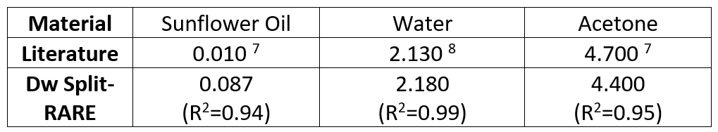
Stejskal-Tanner preparation was used for diffusion sensitization of RARE3. A pair of diffusion gradients was added before and after the first refocusing pulse. A split-echo acquisition was used to deal with the destructive interferences between even and odd echoes, which is due to unknown phase shifts due to the diffusion sensitization module4,5. For initial validation a diffusion phantom was built using a 50ml falcon tube filled with a 5% solution of agarose and three substances with known diffusion properties: sunflower oil, de-ionized water and acetone. Phantom experiments were performed to validate b-value and apparent diffusion coefficient (ADC) map calculation by comparing the experimental data with the literature. Ex-vivo experiments using a perfused rat kidney embedded in agarose and in-vivo experiments with an adult female dark Agouti rat with respiration triggering were performed at a 9.4 Tesla small animal scanner (Bruker Biospec, Ettlingen, Germany). Data was reconstructed offline using custom-made MATLAB code. B-values used for ADC validation were: 0, 200, 400, 600 and 800 s/mm2. Geometric distortions between Dw-SE EPI and Dw Split-RARE were compared and quantified by a center gravity analysis using Dw-Spin Echo image as a reference6. The matrix size and the FOV were adapted to have the same resolution (0.12x0.12x2.0 mm3) in all experiments.Results
Figure 1.a shows phantom images obtained with diffusion-weighting ranging from b=0 s/mm2 to b=800 s/mm2 Figure 1.b depicts the corresponding ADC map. ADC values were 0.087, 2.180 and 4.700s/mm2 for sunflower oil, water and acetone (Table 1). Figure 2 compares high resolution images using Dw-Spin Echo, Dw-SE EPI and Dw Split-RARE for an axial slices of the diffusion phantom, a coronal slices of an ex-vivo rat kidney and a coronal slice of a rat abdomen in-vivo. Severe geometric distortions are present in the Dw SE-EPI images. Figure 3 compares the geometric distortions between Dw-SE EPI and Dw Split-RARE using Dw-Spin Echo image as a reference. Center of gravity analysis revealed a displacement of the center of gravity in pixels with respect to the Dw-Spin Echo reference of (2.4 ± 0.2) for Dw Split-RARE, due to point spread function broadening, (13.1 ± 6.2) for Dw-SE EPI in the phantom and (2.8 ± 1.0) for Dw Split-RARE and (8.6 ± 3.0) for Dw-SE EPI in ex-vivo.Discussion and Conclusion
Diffusion-weighting was successfully implemented in Split-echo RARE. ADC measurements in a diffusion phantom were in line with literature values. Unlike Dw-SE EPI, Dw Split-RARE provided high anatomic fidelity. In in-vivo experiments Dw Split-RARE outperformed Dw-SE EPI by showing both kidneys with no geometric distortions and Dw-Spin Echo by being less prone to motion artifacts due to shorter scan time. To conclude, this study demonstrates the feasibility of Dw Split-RARE at ultrahigh fields for renal imaging. Future in-vivo experiments using (ir)reversible test interventions are needed to evaluate the performance of this approach for the detection of changes in the tubular volume fraction.Acknowledgements
This work was funded by a grant from the German Research Foundation (NI 532/9-1, FOR 1368).References
1. Pohlmann, Acta Physiologica 2013;
2. Pohlmann, Invest. Radiol. 2014;
3. Hennig J, Magn Reson Med 1986;
4. Norris DG, Magn Reson Med, 1992;
5. Schick F, Magn Reson Med,1997;
6. Paul K, Investigative radiology 2015
7. Landolt, Zahlenwert und Funktionen aus Physik, Chemie und Technik, 1969;
8. Deoni, Magn Reson Med, 2004;
Figures
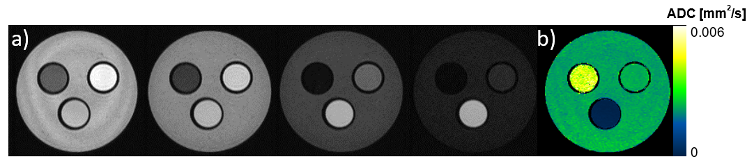
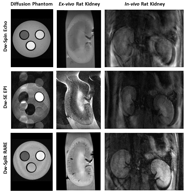
Figure 2: High-resolution images using Dw-Spin Echo, Dw-SE EPI and Dw Split-RARE displaying an axial-view of the diffusion phantom, a coronal-view of an ex-vivo rat kidney and a coronal-view of the abdomen of an adult female dark Agouti rat. Motion artifacts in in-vivo rat kidney are pronounced in Dw-Spin Echo.
Phantom experiment:
Dw-Spin Echo-TE=25ms, TR=2000ms, BW=150000Hz
Dw-SE EPI-TE=76.5ms, TR=2000ms, BW= 350000Hz, segments=4
Dw-Split-RARE-TE=40ms, TR=2000ms, BW=150000Hz, ETL=4
Ex-vivo experiment:
Dw-Spin Echo–TE=24ms, TR=1500ms, BW=150.000Hz
Dw-SE EPI–TE=74.8ms, TR=2000ms, BW=400000Hz, segments=2
Dw-Split-RARE–TE=24ms, TR=1500ms, BW=150000Hz, ETL=4
In-vivo experiment:
Dw-Spin Echo–TE=25ms, TR=2000ms, BW=150000Hz, TA=12min
Dw-SE EPI–TE=28.5ms, TR=2000ms, BW=340900Hz, segments=18, TA=48s
Dw-Split-RARE–TE=21ms, TR=2000ms, BW=150000Hz, ETL=4, TA=3min
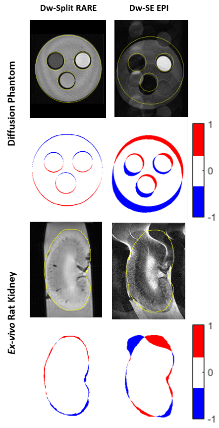
Figure 3: Geometric distortions comparison between Dw Split-RARE and Dw SE-EPI on diffusion phantom and ex-vivo rat kidney using Dw-Spin Echo images as a reference.