3392
The spatial vs. angular resolution trade-off in diffusion MRI explored ex vivo at 9.4T1Harvard-MIT Health Sciences and Technology, Massachusetts Institute of Technology, Cambridge, MA, United States, 2Athinoula A. Martinos Center for Biomedical Imaging, MGH, Charlestown, MA, United States
Synopsis
The time constraints of in vivo diffusion MRI require a compromise to be made between spatial and angular resolution. Given the lack of ground truth on the configuration of human brain connections, determining the optimal operating point along this trade-off is still an open problem. We use high-SNR ex vivo human data at microscopic resolution to study the effect of spatial and angular resolution on dMRI tractography accuracy. Our findings show that voxel size has a much more dramatic effect on tractography reconstruction of challenging white matter configurations than the number of gradient directions.
Purpose
The time constraints of in vivo diffusion MRI (dMRI) require a compromise to be made between spatial resolution and angular resolution. The optimal operating point along this trade-off is not known, and it is likely dependent on the specific white-matter (WM) architecture of interest. The lack of ground truth on the configuration of WM bundles in the human brain makes it challenging to determine optimal acquisition parameters for reconstructing these bundles with dMRI tractography. Here, we take advantage of the high signal-to-noise ratio (SNR) that is feasible only ex-vivo with very long scans at ultra-high field strengths to acquire dMRI data on human brain samples at microscopic resolutions, allowing us to investigate the limits of dMRI performance. We present results from a human WM area with multiple crossing and splitting bundles that we have previously found to cause errors in tractography when compared to chemical tracing in non-human primates1,2. We image it at 9.4T and show the effects that reducing the spatial or angular resolution has on tractography accuracy.Methods
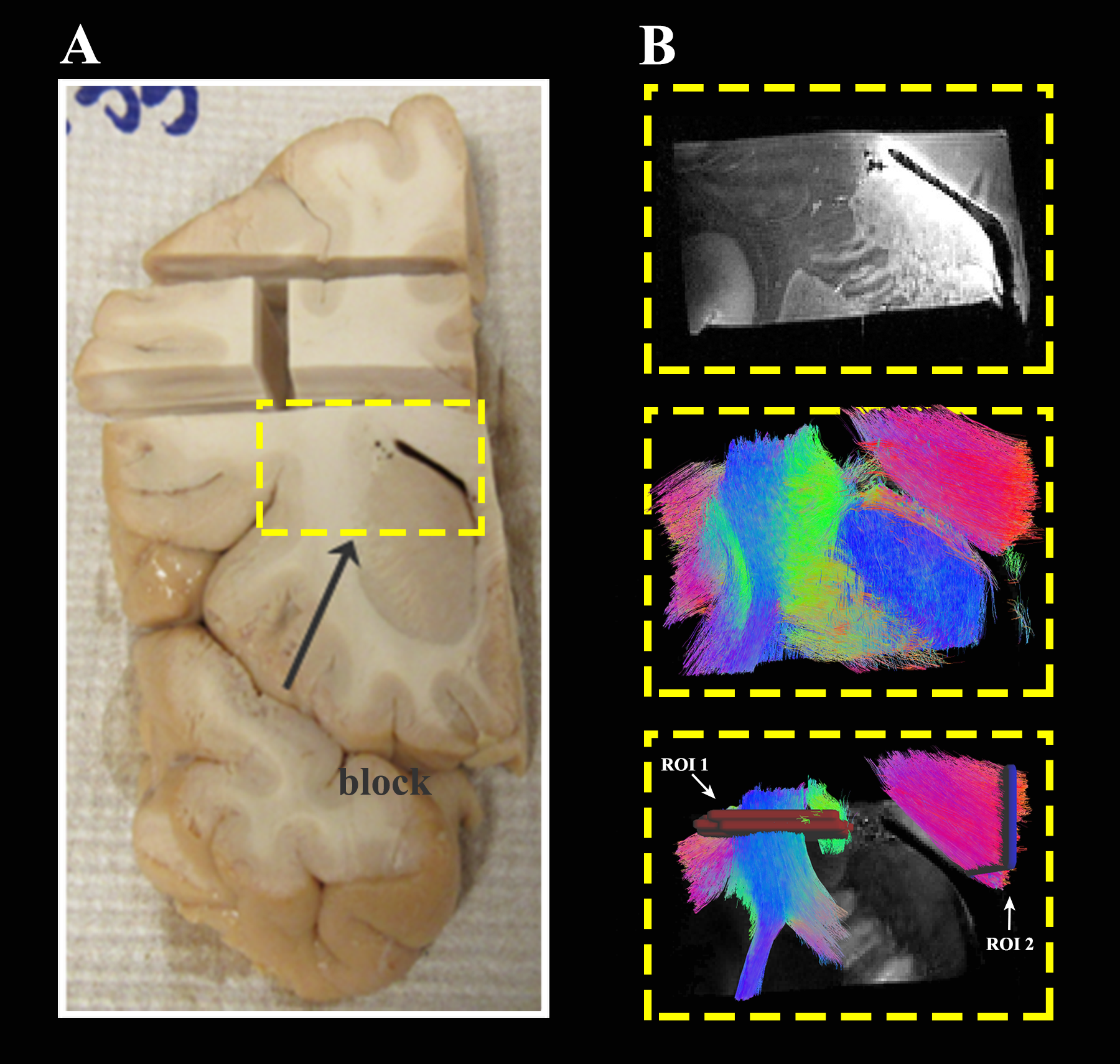
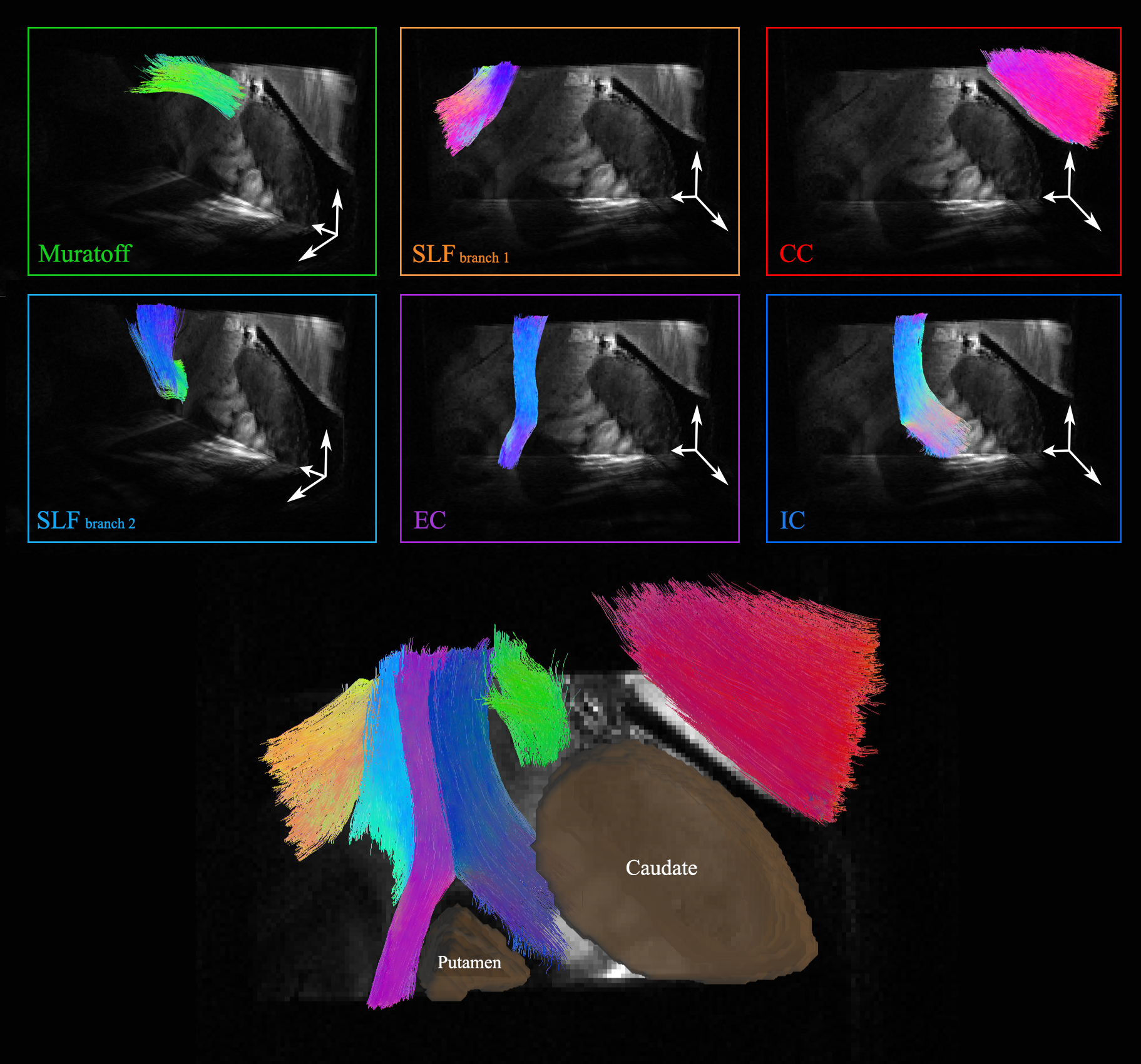
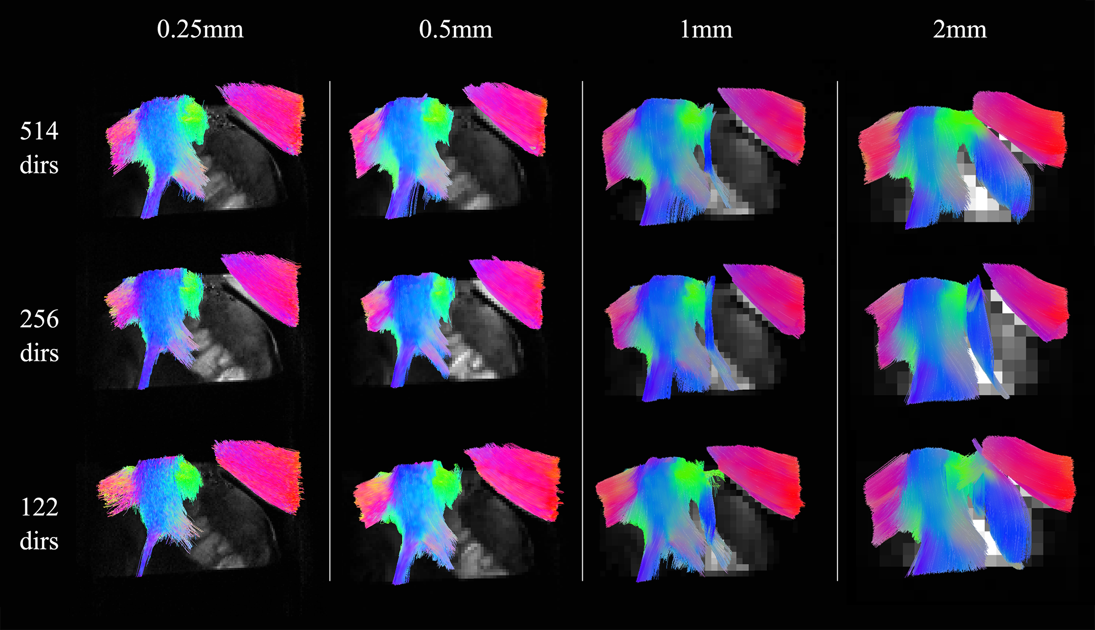
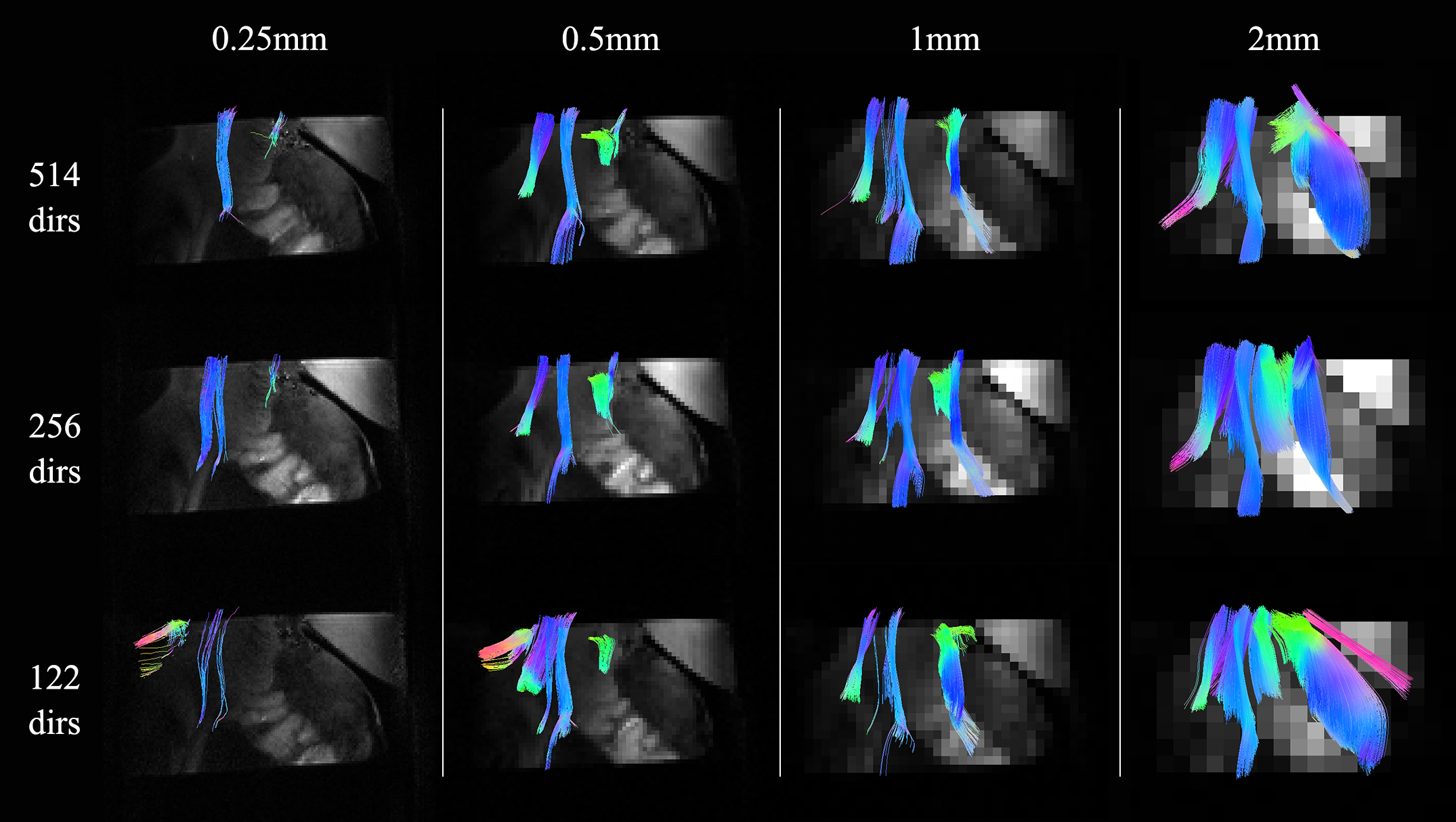
We identify a critical area with crossing and splitting pathways (Figure 1), which we have found to cause errors in prior studies comparing dMRI tractography and chemical tracing in macaques2. We image a 3x2x2cm block of this area from an ex vivo human brain (Figure 1) in a small-bore 9.4T MRI system with maximum gradient=480mT/m. We collect a dMRI dataset using a 2-shot EPI sequence with δ=15ms, Δ=19ms, 514 directions, 0.25mm resolution and bmax=40000s/mm2. From this ultra-high resolution dataset, we generate datasets with varying number of gradient directions (122, 256, and 514) and spatial resolutions (0.25mm, 0.5mm, 1mm, and 2mm). We fit the generalized q-sampling image (GQI) model3 to each dataset and perform deterministic tractography4 with 100000 subvoxel seeds in DSIstudio. Using the two ROIs (ROI1 and ROI2) shown in Figure 1, we select the streamlines we use in our analysis (Figure 3). Then, using additional inclusion and exclusion masks, we isolate, from that subset of streamlines, six unique bundles (Figure 2) in the original dataset and assess how well they can be reconstructed as we reduce the spatial and angular resolution.Results
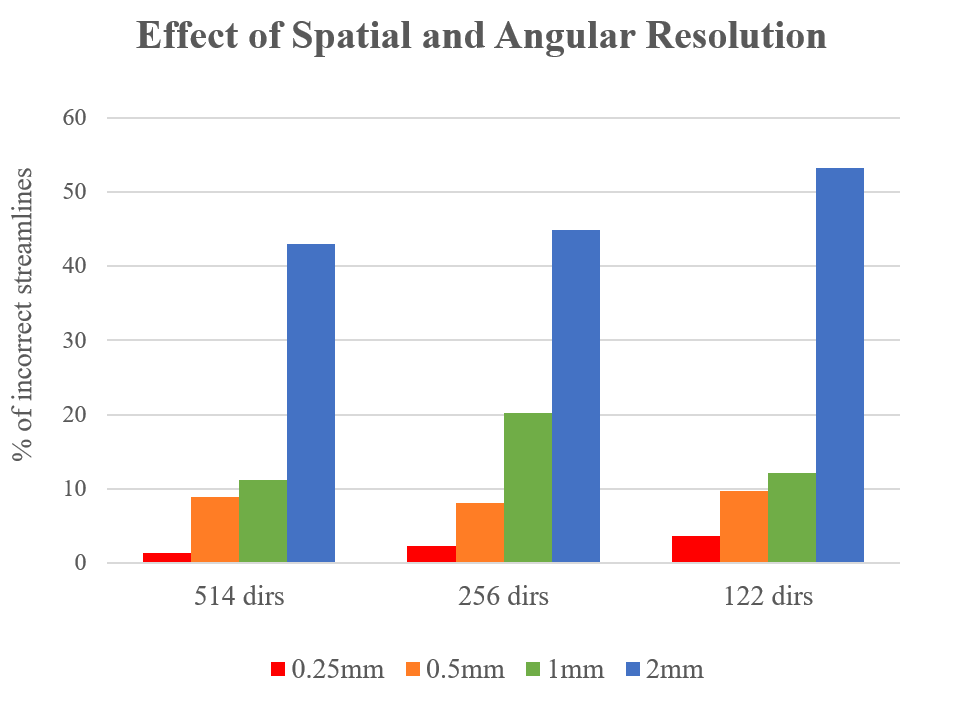
We evaluate tractography accuracy on each dataset by computing the fraction of incorrect tractography streamlines. A streamline is deemed “incorrect” if a) it goes through the inclusion masks for one of the six bundles shown in Fig. 2, but also diverges outside the corresponding bundle, or b) is not neuroanatomically sound1 (e.g. streamlines cutting through the putamen instead of splitting into either extreme capsule or internal capsule). Figure 3 shows the portion of streamlines we used for this analysis and Figure 4 shows which fraction of those streamlines is deemed incorrect. We assess tractography accuracy by calculating the ratio of the number of incorrect streamlines over the total number of streamlines that go through ROIs 1 and 2 of Figure 1; we perform this analysis across datasets with varying spatial resolution and angular resolution (Figure 5).Discussion
Our findings show that spatial resolution has a larger effect on tractography reconstruction than angular resolution. Across all the datasets with each different number of diffusion directions, an increase in voxel size resulted in an increase in incorrect streamlines, with a dramatic accuracy loss at the 2mm resolution that is commonly used in vivo (Fig. 5). Also, the number of errors generally decreased when adding more directions, and this gain was substantial at the 2mm resolution but much less so at higher spatial resolutions.Conclusion
We used ex-vivo human data acquired at an ultra-high field strength to study the effect of spatial and angular resolution on dMRI tractography. Our results indicate that voxel size has a much more dramatic effect than the number of gradient directions on the accuracy of the reconstruction of a challenging WM area in the human brain. These results are specific to the crossing and splitting pathways dorsal to the striatum and the internal capsule, and the analyses methods used here. Our future work will analyze tractography accuracy for pathways from additional areas, as well as under varying SNR levels. Ultimately, our goal is to determine whether there is a trade-off between SNR, spatial and angular resolution that is feasible in vivo and that yields satisfactory tractography performance.Acknowledgements
This research was carried out in whole or in part at the Athinoula A. Martinos Center for Biomedical Imaging at the Massachusetts General Hospital, and was funded by NIH P50 MH106435-01, R01 EB021265, R01 5RO1MH04557325, NBIB P41EB015896 and the NIH Blueprint for Neuroscience Research (T90DA022759/R90DA023427). This work also involved the use of instrumentation supported by the NIH Shared Instrumentation Grant Program and/or High-End Instrumentation Grant Program; specifically, grant number(s) S10RR016811, S10RR023401, S10RR019307, S10RR019254, S10RR023043.References
1. Schmahmann, J. D. & Pandya, D. N. Fiber Pathways of the Brain. New York Oxford University Press 1, (2006).
2. Grisot, G. et al. Optimization of acquisition parameters for diffusion MRI using chemical tracing. ISMRM proceedings, (2016).
3. Yeh, F. C., Wedeen, V. J. & Tseng, W. Y. I. Generalized q-sampling imaging. IEEE Trans. Med. Imaging 29, 1626–1635 (2010).
4. Yeh, F. C., Verstynen, T. D., Wang, Y., Fernández-Miranda, J. C. & Tseng, W. Y. I. Deterministic diffusion fiber tracking improved by quantitative anisotropy. PLoS One 8, (2013).
Figures