3365
Comprehensive analysis of the predictors of microvessel invasion in hepatocellular carcinoma: Diffusion kurtosis imaging (DKI) combined with radiological and clinical factors.1Radiology department, zhongshan Hospital, Shanghai, People's Republic of China, 2MR Application Developmen, Siemens Healthcare, Erlangen, Germany, 3Siemens Shenzhen Magnetic Resonance Ltd, shenzhen, People's Republic of China, 4MR Collaboration NE Asia, Siemens Healthcare, shanghai, People's Republic of China
Synopsis
Diffusion Kurtosis Imaging (DKI) maps, preoperative radiological features and clinical-pathologic findings were calculated to assess their diagnostic accuracy for microvascular invasion (MVI) of hepatocellular carcinoma (HCC) in patients who were undergoing curative liver resection. Multivariate regression analysis was performed to identify independent predictive factors for MVI. The study shows that Mean Kurtosis (MK), non-smooth margin, peritumoral enhancement and incomplete radiological capsule suggest a high probability of microvessel invasion of HCC. Multivariate analysis confirmed that MK and capsule integrity show statistical significance correlation with MVI. In conclusion, MK and capsule appearance might be the predictors for MVI of primary hepatocellular carcinoma.
Introduction and purpose
The
diagnosis of hepatocellular carcinoma (HCC) at a curable stage can be based on
current imaging techniques and/or biopsy. However, the high recurrence rate at
5 years either after resection or transplantation still remains a challenge in
HCC management [1]. There is a lack of consensus on the definition of
microvessel vascular invasion (MVI) and the features with the strongest
prognostic association. Jensen et al proposed a non-Gaussian diffusion-weighted
model called diffusion kurtosis imaging (DKI) in 2005, which might provide more
accurate information about water diffusion and shows substantially higher
sensitivity for tumor detection [2]. This study aimed to evaluate the accuracy
of DKI and conventional ADC, together with radiological features and clinical
findings in the prediction of MVI in HCC.
Materials and Methods
Seventy-eight patients with eighty-five lesions underwent MR exams with a 1.5T MR scanner (MAGNETOM Aera, Siemens, Erlangen, Germany). A prototype single-shot spin-echo echo-planar DW imaging sequence was used to acquire the DKI data under free-breathing. Parameters were: b values = 0, 200, 500, 1000, 1500, 2000 sec/mm2 (with average of 1, 1, 2, 2, 3, 4), TR =8000ms, TE = 63ms, FOV = 308x380mm2, scan matrix = 128x80, slice thickness = 5mm, transversal orientation with coverage of the whole liver, total scan time = 1min 59sec. Mean kurtosis (MK) maps, mean diffusion (MD) maps of DKI model, and conventional ADC maps were calculated by using a prototype software (Body Diffusion Toolbox, Siemens Healthcare, Erlangen, Germany). The radiological features were retrieved from our institutional picture archiving system ( PACS; Pathspeed , GE Medical systems Integrated imaging Solutions, Prospect, IL, USA). The histopathologic diagnosis of MVI of primary HCC was confirmed by surgical resection.Results
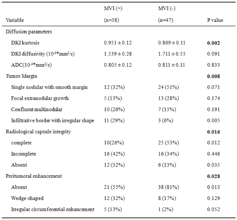
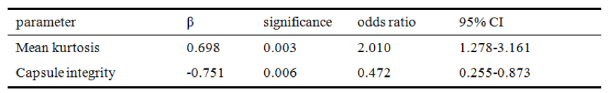
The univariate analysis of imaging parameters is given in Table 1. The MK values were significantly higher in the MVI-positive lesions than in the negative lesions (0.951 ± 0.12 vs 0.869 ± 0.11. P=0.002). Tumor margin (p=0.008), radiological capsule (p=0.016) and peritumoral enhancement (p=0.028) were also associated with MVI. In the multivariate analysis (logistic regression analysis, Table 2), MK showed statistical significance (odds ratio 2.010, β 0.698), suggesting a high probability of MVI of HCC. Capsule integrity also showed statistical significance (odds ratio 0.472, β -0.751), suggesting the integrity of the capsule may be the protective factor for MVI of HCC.Discussion
MVI is an expression of aggressive histological feature that is considered as one of the most common risk factors for recurrence after resection and worse prognosis for HCC. Several studies have proposed some preoperative imaging features and clinical factors for prediction of MVI in HCC patients [3,4]. Furthermore, previous studies on HCC [5-6] found that DKI showed higher accuracy than conventional DWI for characterizing tumor tissues. The multivariate analysis in our study showed MK and capsule appearance were significant independent risk factors for predicting MVI. This might be due to the ability of DKI for reflecting the heterogeneity and irregularity of cellular microstructure. Hence, MK has potential value as surrogate marker for predicting MVI of HCC.Acknowledgements
No acknowledgement found.References
[1] Bruix J, Sherman M. Hepatology 2005;42:1208–36.
[2] Jensen JH, et al. Magn ResonMed2005;53(6):1432–1440.
[3] Kaibori M, et al. J Surg Oncol 2010;102:462–8.
[4] Kim BK, et al. J Surg Oncol 2008;97:246–52.
[5] Goshima S, et al. AJR Am J Roentgenol 2015; 204:W543–549.
[6] Rosenkrantz AB, et al. Magn Reson Imaging 2012;30(10):1534–1540.
Figures