3355
Evidence for Cross-Term Effects in Double Diffusion Encoding Experiments of Human Cortical Gray Matter1Department of Systems Neuroscience, University Medical Center Hamburg-Eppendorf, Hamburg, Germany, 2Neuroimage Nord, University Medical Centers Hamburg-Kiel-Lübeck, Hamburg-Kiel-Lübeck, Germany
Synopsis
Double diffusion encoding (DDE) experiments with two weighting periods applied successively in the same acquisition are a promising tool to investigate microscopic tissue properties, e.g. the cell eccentricity and the related diffusion anisotropy on a microscopic scale. Recent experiments detected the signal pattern typical for microscopic diffusion anisotropy in human cortical gray matter in vivo but were hampered by an additional signal modulation that could be related to field inhomogeneities near the skull. In this study, cross-term-compensated DDE experiments are performed to investigate the effect of field inhomogeneities on the detection of the microscopic diffusion anisotropy.
Introduction
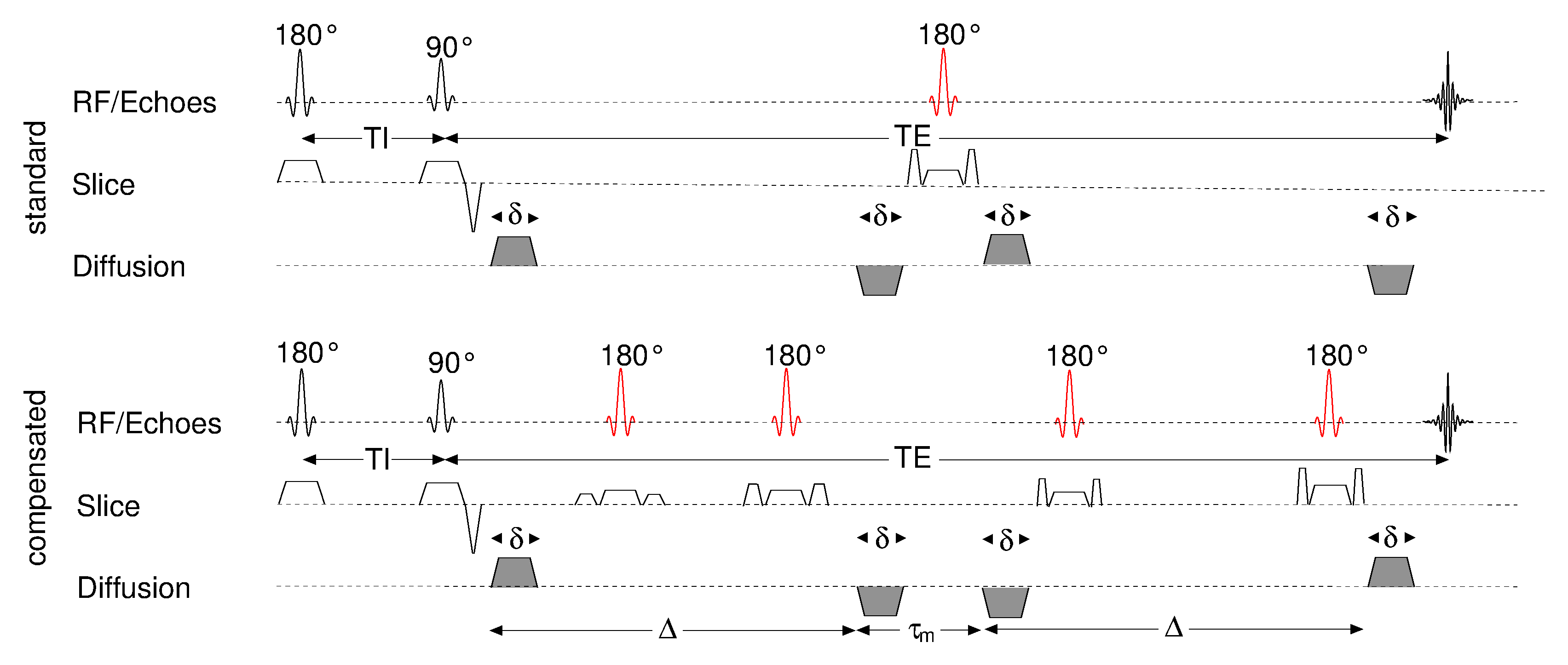
Double diffusion encoding (DDE) or double wave vector experiments (DWV) [1-3] using two diffusion weighting periods applied successively in a single acquisition (Fig. 1) offer access to microscopic tissue properties [1-4]. The typical fingerprint of the angular DDE experiment at long mixing times τm for eccentric cells or microscopic diffusion anisotropy is a “W”-shaped signal modulation with maxima for parallel/anti-parallel and minima for orthogonal weightings [1]. This anisotropy effect has even been detected in a human brain white matter region-of-interest that appeared isotropic in diffusion tensor imaging (DTI) [5]. A similar signal difference between parallel/anti-parallel and orthogonal weightings could be detected in human cortical gray matter [6]. However, the measurements were often hampered by an additional, “U”-shaped signal modulation. It has been hypothesized that this modulation could arise from cross- terms with magnetic field inhomogeneities close to the skull [6]. In this study, experiments with cross-term compensated DDE measurements were performed that provide evidence for this hypothesis.Methods
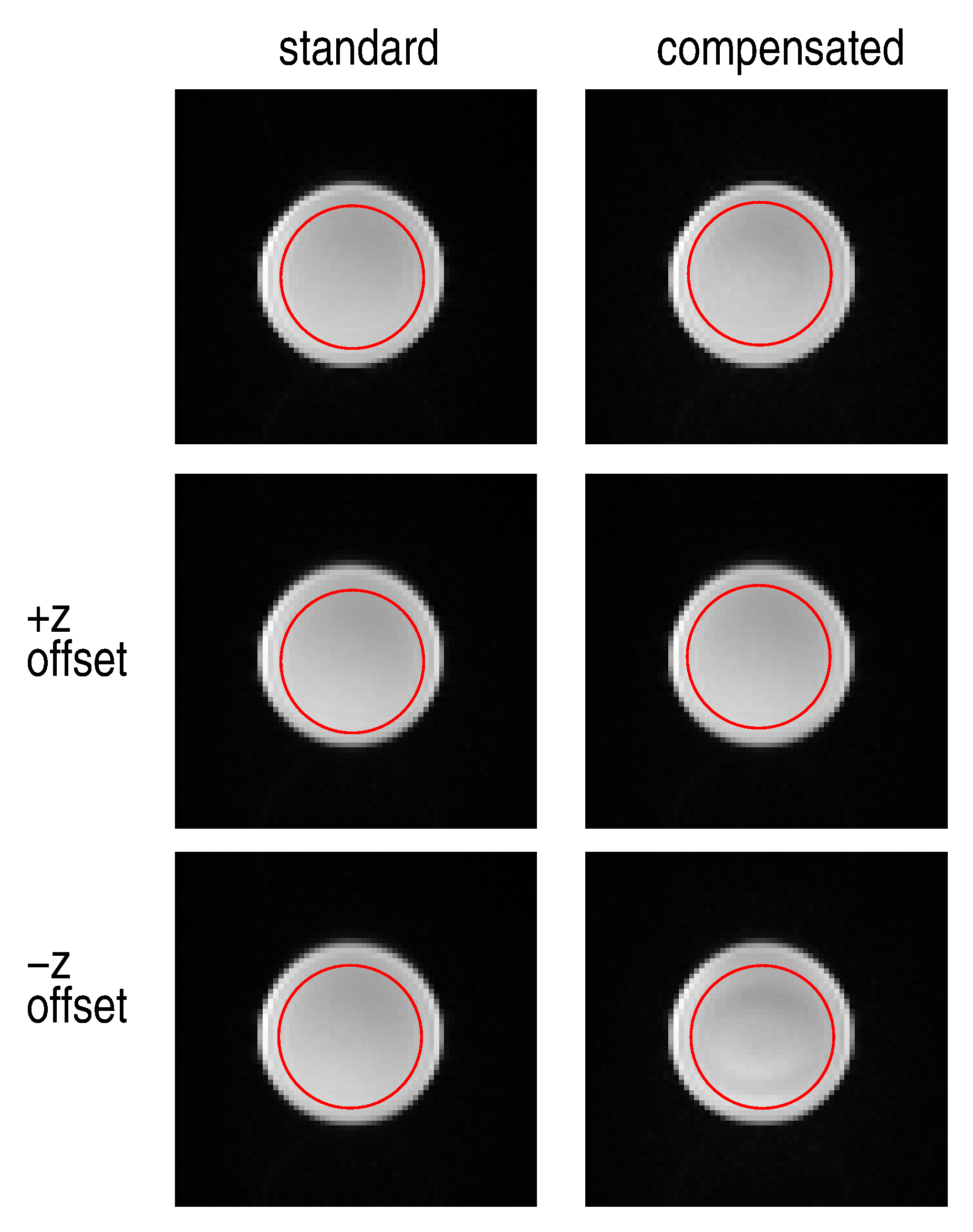
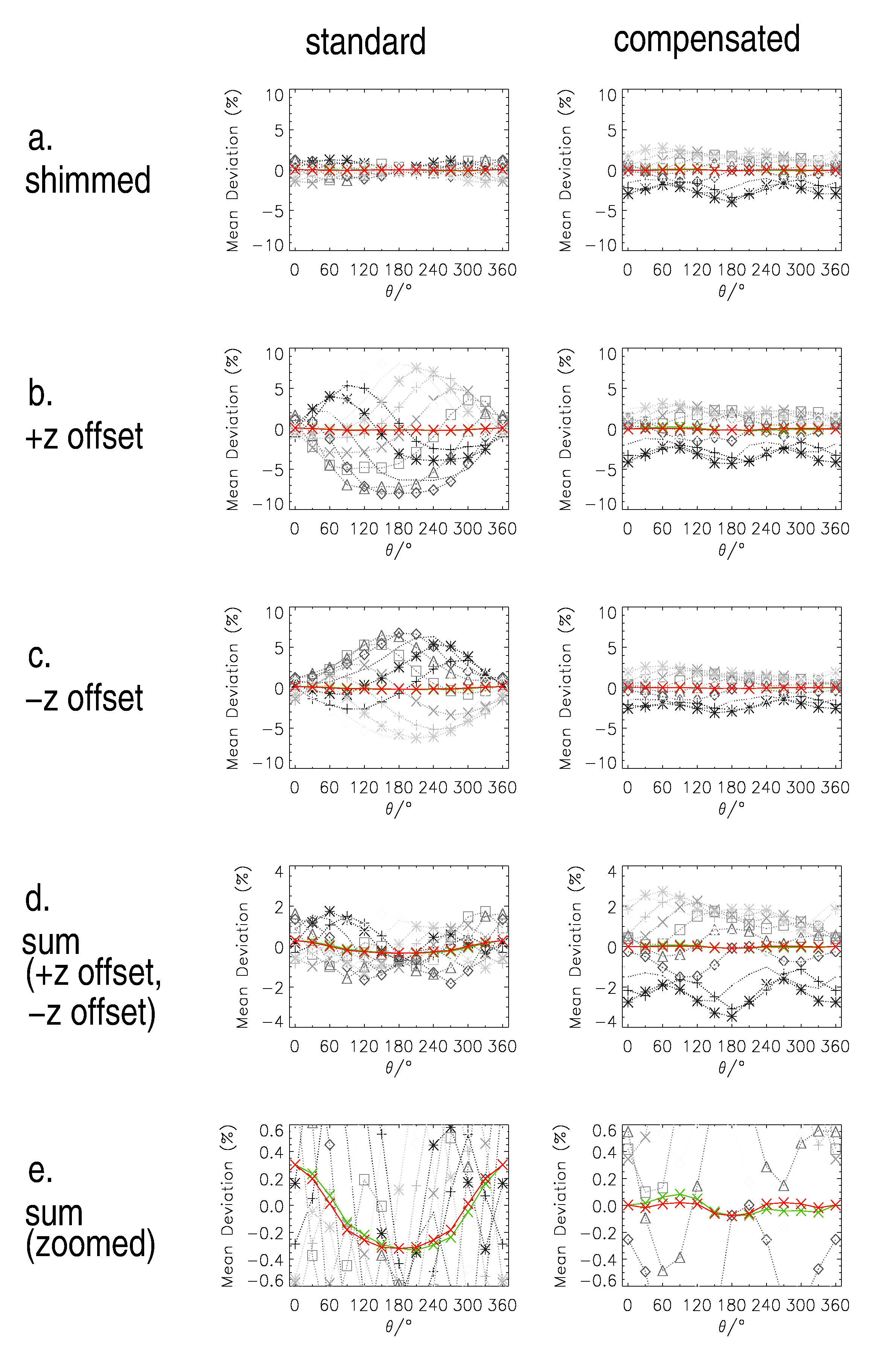
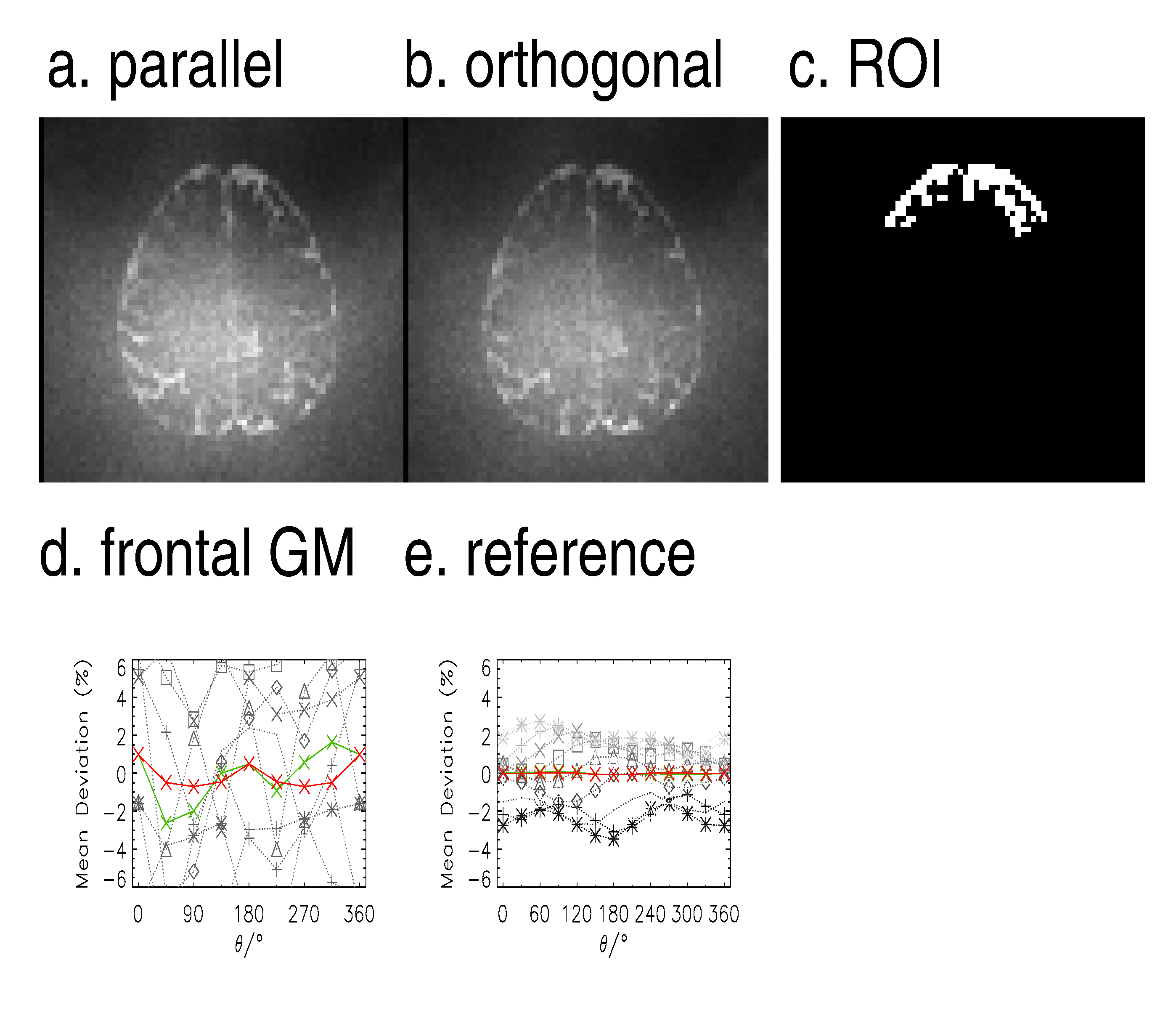
DDE experiments were performed on a 3T whole-body MR system (TIM Trio, Siemens Healthcare) on healthy volunteers after their informed consent was obtained. A dodecane (C12H26) phantom with purely isotropic diffusion was used as a reference. For the latter, a misadjustment of the linear shim term in z (slice direction) was used to simulate the effect of field inhomogeneities. Thereby, two measurements with two polarities of the misadjustment were performed that could be averaged to mimic the effect of variable field inhomogeneities in a large ROI. The basic pulse sequences are sketched in Fig. 1. An inversion pulse was applied prior to the imaging sequence with a recovery time of TI = 630 ms that effectively nulls brain white matter signals in vivo. Two DDE preparations with long mixing times were considered: (i) the standard preparation with a single refocusing RF pulse in the middle of the mixing time interval between the two weightings (“standard”) and (ii) a preparation involving two refocusing RF pulses per diffusion weighting that nulls cross-terms with linear field gradients for each of the diffusion weightings (“compensated”). The image readout was performed as in a standard echo-planar imaging sequence (Fig. 1) with voxel sizes of 3.0 x 3.0 x 3.0 mm3 (phantom) and 4.0 x 4.0 x 3.0 mm3 (in vivo) resolution (TE/TR = 265 ms /6.5 s). Only a single transverse slice was measured to minimize table vibration effects. Both preparations had identical timings for the two diffusion-weighting periods that were applied with a b value of 500 s mm-2 each, a diffusion time δ of 8 ms, a mixing time τm of 18 ms, and a gradient pulse duration Δ of 90 ms (compare [7]). All 144 combinations of 12 diffusion encoding directions sampling a circle in steps of 30° were applied for the xz-coordinate plane that involves the misadjusted linear shim term in the phantom experiments. Having one image without diffusion weighting, the total acquisition time was 14.5 min. The MR signal variations with the angle θ in a region-of-interest in the phantom (cf. Fig. 2) and in a frontal gray matter regions in vivo (cf. Fig. 4) were plotted individually, averaged over all 12 circles, and geometrically averaged with the anti-podal setup, respectively.Results and Discussion
The MR individual signal curves for the fluid phantom shown in Fig. 3 reveal variable modulations with the angle θ, inter alia due to cross-terms with spoiler and slice selection pulses, but the geometrically averaged curves (green) as well as the symmetrized averaged (red curves) show almost flat lines in the absence of field inhomogeneities (Fig. 3a) as expected. However, with field inhomogeneities involved (Fig. 3b-e), the averaged curve for the standard preparation shows a “U”-shaped pattern very similar to that observed in previous experiments in human cortical gray matter [6]. In contrast, the compensated preparation shows a significantly reduced modulation with very similar signals for parallel and antiparallel weightings which seems to indicate that field inhomogeneities can induce a “U”-shaped signal curve. In Fig. 4, the signal curves for the phantom and in vivo data obtained with the compensated DDE preparation are compared. In frontal cortical gray matter an almost undisturbed “W”-shaped curve is observed that is expected in the presence of microscopic diffusion anisotropy. This modulation is much larger than the one present in the phantom underpinning that the anisotropy effect is related to the tissue.Conclusion
DDE-based measurements of the microscopic diffusion anisotropy in human cortical gray matter may be hampered by cross-terms with field inhomogeneities but their influence can be minimized with a correspondingly adjusted preparation.Acknowledgements
No acknowledgement found.References
[1] Mitra PP, Multiple wave-vector extensions of the NMR pulsed-field-gradient spin-echo diffusion, Phys. Rev. B 51, 15074 (1995)
[2] Shemesh N, Conventions and nomenclature for double diffusion encoding (DDE) NMR and MRI, Magn. Reson. Med. 75, 82 (2015)
[3] Cheng Y, Multiple scattering by NMR, J. Am. Chem. Soc. 121, 7935 (1999)
[4] Özarslan E, Compartment shape anisotropy CSA revealed by double pulsed field gradient MR, J. Magn. Reson. 199, 56 (2009)
[5] Lawrenz M, Double-wave-vector diffusion-weighted imaging reveals microscopic diffusion anisotropy in the living human brain, Magn. Reson. Med. 69, 1072 (2013)
[6] Lawrenz M, Detection of Microscopic Diffusion Anisotropy in Human Brain Cortical Gray Matter in Vivo with Double Diffusion Encoding, Proc. ISMRM 2016, p. 212
[7] Lawrenz M, Mapping measures of microscopic diffusion anisotropy in human brain white matter in vivo with double-wave-vector diffusion-weighted imaging, Magn. Reson. Med. 73, 773 (2015)
Figures