3354
Reducing Distortion in DWI Acquisitions of Prostate Scans1Mayo Clinic, Rochester, MN, United States
Synopsis
Diffusion images obtained with spin echo – echo planar imaging (SE-EPI) can be acquired quickly and efficiently. In prostate applications areas of susceptibility can cause image distortion. The purpose of this work is to address these distortions with the use of multi-shot EPI techniques. A previously defined term, pseudo-gradient, is shown to be a useful analytical tool to explain and guide in the choice of acquisition parameters. Phantom and in-vivo results show the benefits of multi-shot scans to reduce distortions.
PURPOSE
Diffusion images obtained with spin echo – echo planar imaging (SE-EPI) can be acquired quickly and efficiently. In prostate applications areas of susceptibility can cause image distortion. To address this, a SE-EPI based acquisition called field-of-view (FOV) optimized and constrained undistorted single-shot (FOCUS) 1 sequence has been developed to reduce geometric distortions. This has recently been adapted for prostate DWI 2,3. However, distortions remain an issue in prostate SE-EPI due to the air-filled rectum and continue to degrade image quality. The purpose of this work is to further address this with the use of multi-shot EPI techniques.
Methods
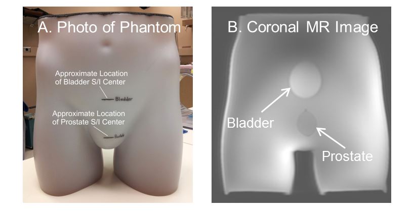
The sensitivity of EPI to distortion can be expressed in terms of the pseudo-gradient J 1 $$J = \frac{2\pi\times R_{y}\times m}{\gamma\times FOV_{y}\times T}$$ where $$$R_{y}$$$ is acceleration along the Y direction, m is the number of shots for multi-shot acquisition, and T is the time between echo centers. As J decreases, the off-resonance worsens. Conversely, as J increases the resulting geometric distortion will decrease. In multi-shot EPI the effective time between echo centers in the resultant fully sampled k-space is reduced proportionally by the number of shots m. With a decrease in T, the pseudo-gradient will increase and reduce off-resonance effects and geometric distortions. In this work a FOCUS pulse sequence was modified to allow for multi-shot acquisition. For controlled phantom studies a bovine gelatin-filled pelvic phantom seen in Figure 1 was developed to examine this relationship. The phantom contains an air-filled cavity simulating the rectum in close proximity to a simulated prostate and bladder inclusions. The multi-shot FOCUS DWI EPI sequence was also studied in in vivo.
RESULTS
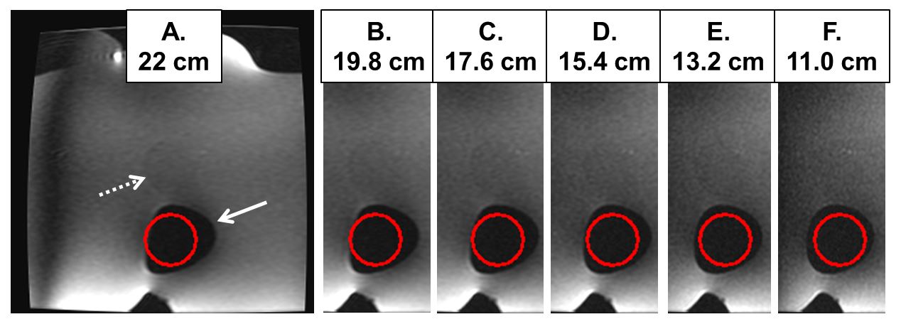
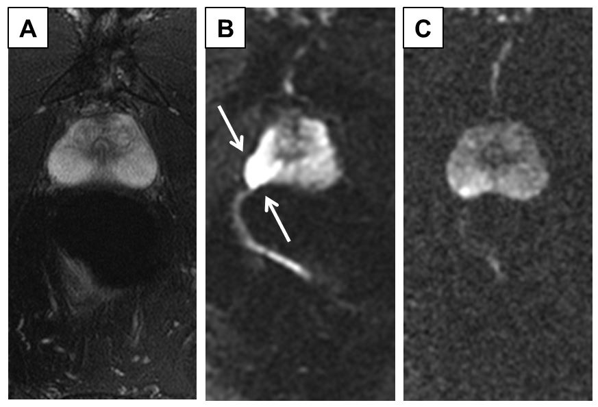
As a starting point the phantom was imaged with a limited-FOV sequence using progressively smaller phase (L/R) FOVY, Figure 2. Due to the susceptibility difference in the abdominal phantom between prostate and air, off-resonance effects are created. Note the progressive improvement in the fidelity of portrayal of the ellipsoidal-shaped dark rectum with reduced FOV. This improvement can be explained by observing that a reduction in FOVY will increase the pseudo-gradient J and thus result in lower geometric distortion. Diffusion single-shot and two-shot images were obtained using a diffusion weighted imaging (DWI) FOCUS acquisition as seen in Figure 3. The number of averages was modified to keep the scan time constant for the two scans. Note the geometric distortion in Figure 3B due to the off-resonance of the tissue-air interface with the rectum as well as the signal pileup within the prostate. The two-shot acquisition, Figure 3C, shows reduced distortion and more uniform image intensity.
DISCUSSION
Geometric distortion due to off-resonance effects remain a continuing issue in prostate DWI imaging. We have shown that the pseudo-gradient can be a useful analytical tool to explain and guide in the choice of acquisition parameters. While multi-shot FOCUS can be used to reduce distortions, the resulting scan time increases and ideally is accounted for. For example, the number of sample averages can be reduced at the cost of lowering signal to noise ratio (SNR) in the multi-shot images. In future work we will look to optimize b-value sampling strategies and ADC calculations to maximize use of the available SNR.Acknowledgements
No acknowledgement found.References
1 Jeong EK, Kim SE, Guo J, et al. High-resolution DTI with 2D interleaved multislice reduced FOV single-shot diffusion-weighted EPI (2D ss-rFOVDWEPI). Magn Reson Med 2005;54:1575–79. 2 Thierfelder KM, Scherr MK, Notohamiprodjo M, Weiss J, Dietrich O, Mueller-Lisse UG, Pfeuffer J, Nikolaou K, Theisen D. Diffusion-weighted MRI of the prostate: advantage of zoomed EPI with parallel-transmit-accelerated 2D-selective excitation imaging. Eur J Radiol 2014;24:3233-3241. 3 Korn N, Kurhanewicz J, Banerjee S, Starobinets O, Saritas E, Noworolski S. Reduced-FOV excitation decreases susceptibility artifact in diffusion-weighted MRI with endorectal coil for prostate cancer detection. Magn Reson Img 2015;33:56-62. 4 Farzaneh F, Riederer SJ, Pelc NJ. Analysis of T2 limitations and off-resonance effects on spatial resolution and artifacts in echo-planar imaging. Magn Reson Med 1990;14:123-139.
Figures