3351
Correcting a slice distortion artifact in the multiband diffusion images1University of Southern California, Los Angeles, CA, United States
Synopsis
The diffusion weighted images acquired with the multiband sequence or the Lifespan protocols shows a type of slice distortion artifact. We find that this artifact is caused by the eddy currents, which can be induced by the diffusion gradient associated with either current DW image or the previous DW images. The artifact can be corrected by further tuning the compensation circuit in the MR hardware, or by a correction algorithm which includes the diffusion gradients from the current and previous DW images.
Introduction
Diffusion tensor imaging (DTI) or high angular resolution diffusion imaging (HARDI) has become a popular tool to provide information about the intrinsic architecture of white matter in the human brain. However, because most of the HARDI techniques require high to ultrahigh diffusion sensitizing gradients, the capability of HARDI to provide valid and reliable information about tissue structures can be affected adversely by eddy current artifacts [1]. In echo planar images, usually used to acquire diffusion-weighted images (DWI), eddy currents produce significant distortions in the phase-encoding direction because of the relatively low bandwidth in that direction and the large changes in diffusion gradients. The recently developed multilband slice-accelerated technique can provide high angular resolution for measuring the diffusion signals in shorter time length than conventional EPI sequence [2], especially in Human Connectome Lifespan projects. However, how the eddy current specially affects the multiband diffusion images or whether the multiband acquisition is special on the characters of eddy current is still under investigation.Methods and Results
A. Inspection
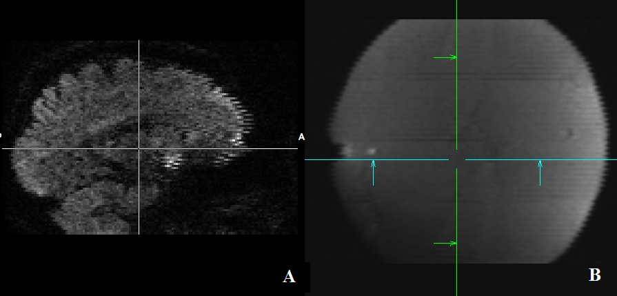
One fBIRN phantom and two adults with no history of neurological disease were scanned on a Siemens 3T Trio Prisma MRI system (Siemens Healthcare, Erlangen, Germany) at our institute. HARDI data were acquired using multiband DWI sequence with the following parameters as in the Lifespan projects: repetition time (TR) 3222 msec, echo time (TE) 89.2 msec, 197 diffusion gradient directions, 92 axial slices for whole brain coverage, MB factor as 4, and maximum b value as 2000 s/mm^2. As in the result images (Figure 1), an artifact shows up as misalignment between slices in both the diffusion weighted images and b0 images in both human and phantom data.
B. Diagnosis
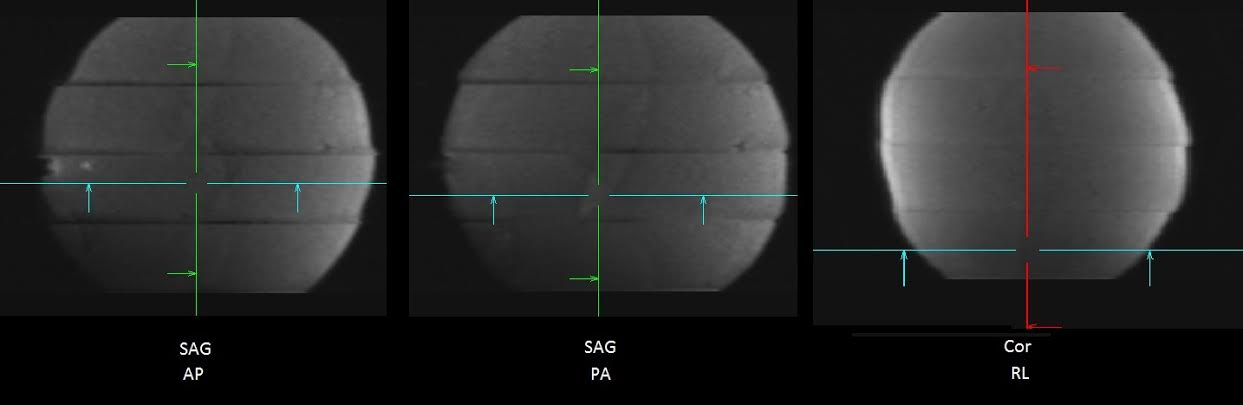
To diagnose the possible reason of this artifact, we change the slice order from interleaved into ascending, and switch the phasing encoding direction into AP, PA and RL in the DWI scans on the fBIRN phantom. The results show that the PA and AP phase encodings reverse the slice stretch and shrink at the sagittal view, and RL phase encoding makes the slice shift at the coronal view (Figure 2). It is a typical eddy current effect. However, this kind of eddy current artifact is rarely seen on the b0 images in the diffusion scans and in the conventional EPI images. The further diagnosis on the eddy currents on the MRI is done on the next.
C. Hardware correction
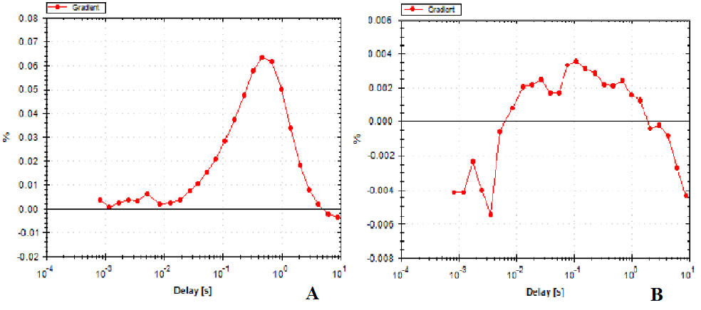
Using Siemens service hardware, we map the eddy current along the time axis (Figure 3A). We found the typical compensation of eddy current mainly focused on the time range of 2-100ms, but the amplitude of eddy current around 1-4 seconds is still large. This time range is close to the TR (3222 ms) used in the Lifespan protocol. The super short TR and super high diffusion gradient strength in this MB protocol make it possible that the eddy current affects different images across TR. That is why the b0 image in the Lifespan protocol displays this artifact. Before multiband sequence is available, most diffusion scans of the whole brain have TR much longer by a few seconds, so this eddy current effect from previous volume is very low in the conventional EPI images. After our re-tuning, the eddy current after the specified time range (1-4s) is also reduced (Figure 3B). Therefore this artifact is deducted significantly in the new images (Figure 4B).
D. Software correction
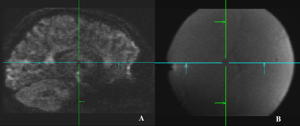
To correct the diffusion weighted images which already has this artifact, we apply the algorithm for correcting the eddy current distortions using the known diffusion gradient strengths and directions, which was published before [1]. However, because the the distortion in one image is not only affected by the current diffusion gradient but also by the previous diffusion gradient. Thus the algorithm should include the diffusion gradient associated with the previous diffusion weighted volume. After correction by this way, the artifact is reduced to the minimal level (Figure 4A).
Conclusion
The slice distortion shown in the diffusion weighted images acquired with the multiband sequence or the Lifespan protocols is caused by the eddy currents, which can be induced by the diffusion gradient associated with either current DW image or the previous DW images. It can be corrected by further tuning the hardware compensation, or by a correction algorithm which includes the diffusion gradients from the previous DW image.Acknowledgements
No acknowledgement found.References
[1] Zhuang, et al., JMRI: 1460, 2013.
[2] Feinberg, et al., PloS ONE: e15710, 2010.
Figures