3348
Simultaneous multi-slice (SMS) echo planar imaging (EPI); which combination of parameters is clinically most efficient?1Kyoto Prefectural University of Medicine, Kyoto, Japan, 2Siemens Japan K. K., 3Kyoto Prefectural University of Medicine Hospital, 4Siemens Medical Solutions USA
Synopsis
To find clinically feasible combinations of parameter for simultaneous multi-slice echo planar imaging (SMS-EPI) acquisition diffusion tensor imaging (DTI), we investigated the combinations of SMS factors and the amounts of inter-slice image shift to find which one can provide better signal-to-noise ratio (SNR) by two different head coils (20ch and 32ch), respectively. The SMS factors 2 and 3 with imaging shifts (>1/2) on 32ch judged clinically feasible and can be substituted on conventional DTI. The SMS factor 2 with imaging shifts 0, 1/2, 1/4 on 20ch head coil judged clinically feasible.
PURPOSE
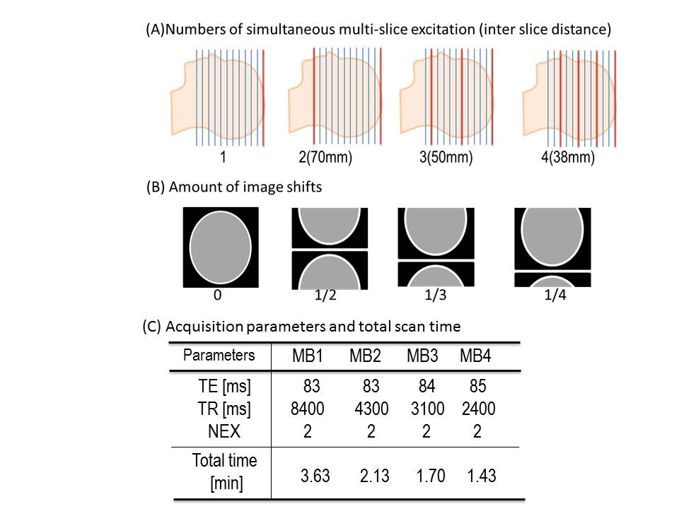
Simultaneous multi-slice (SMS) echo planar imaging (EPI) acquisition appears useful for clinical diffusion tensor imaging (DTI) to reduce acquisition time dramatically. However, the optimal combination of SMS-EPI parameters for clinical DTI remains unclear. There are three parameters available for optimizing image quality for SMS-EPI DTI: the number of slices excited and acquired simultaneously (SMS factor); the amount of inter-slice image shift (one-dimensional CAIPIRINHA[1]); and the distance between slices excited simultaneously (Fig. 1). We investigated combinations of these parameters to find which one can provide better signal-to-noise ratio (SNR), and diffusion metrics (ADC and FA) for two different head coils (20ch and 32ch), respectively.METHODS
Subject: This study was approved by the Ethics Committee at our Institution. The subject consisted of one healthy, 47-year-old, male volunteer.
Data acquisition: MRI examinations were performed on a clinical 3 T whole-body scanner (MAGNETOM Skyra, Siemens Healthcare, Erlangen, Germany) with two different head coils (20ch and 32ch). Scan parameters were : in-plane field of view (FOV) : 240 x 216 mm, acquisition matrix : 120 x 108, b value = 1,000 s/mm2, 20 diffusion directions were acquired. DTI was acquired using a prototype SMS-EPI sequence with inter slice image shifts (shift 0, 1/2, 1/3 and 1/4, Fig.1B), called “blipped CAIPIRINHA [1]”. A total of 70, 75 and 76 2-mm-thick slices were obtained without inter-slice gaps with SMS factor 1, 2, 3, and 4, respectively. The inter slice distance for SMS factor 2, 3, and 4 was 70, 50, 38mm, respectively (Fig.1A). Repetition time, echo time, NEX, and total scan time are summarized in Fig.1C.
DTI analysis: DTI analysis was performed in Diffusion toolkit [2] and diffusion metrics (mean DWI, ADC and FA) were calculated.
SNR comparison: We obtained a registered brain parcelation map [3] by LDDMM on Diffeomap [4]. Signal to noise ratio (SNR) was calculated within the each parcel on the parcelation map by differential method between two independent DTIs [5]. The relative SNR to conventional DTI (SMS1) was compared on each parcel at different SMS factors and different inter slice image shifts.
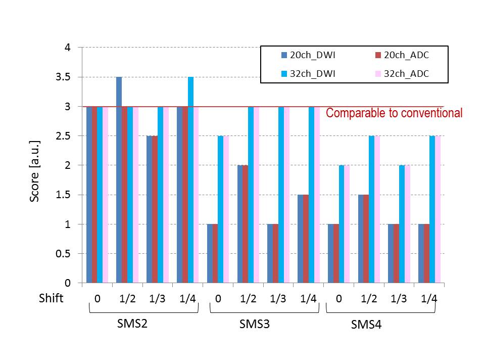
Evaluations of image quality: Image quality of ADC and FA were evaluated by comparison to conventional DTI. To compare both ADC and FA to conventional DTI, we employed tract based statistics by TrackVis [2] and calculated relative ADC and FA. To evaluate clinical feasibility, we compared image quality of conventional DTI and SMS-EPI DTI (compared to conventional, SMS-EPI DTI is much worse (1) / slightly worse (2) / comparable to (3) / slightly better (4) / much better (5)) on DWI and ADC by two experienced neuroradiologists (J.T. and M.Y.).
RESULTS AND DISCUSSION
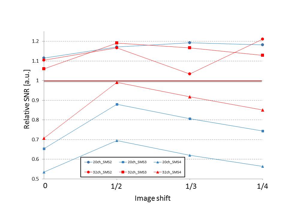
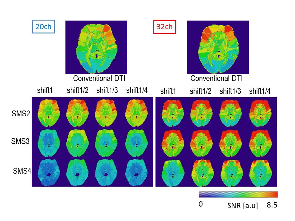
Effect of SMS factor on SNR: Figure 2 shows the relative SNRs of the two head coils. On the 20ch head coil, only SMS2 showed larger SNR than conventional DTI. On the other hand, both SMS2 and SMS3 showed larger SNR than conventional DTI on the 32ch head coil. Figure 3 shows parceled SNR maps of both 20ch and 32ch head coils, clearly showing that SNR is better with the 32ch head coil than with the 20ch coil.
Effect of inter slice shift on SNR: In Figure 2, the horizontal axis shows the amount of interslice image shift. With all amounts of image shift, SNRs were better than without image shifting. In most SMS factor cases, 1/2 shift was effective to achieve better SNR compared to the conventional method.
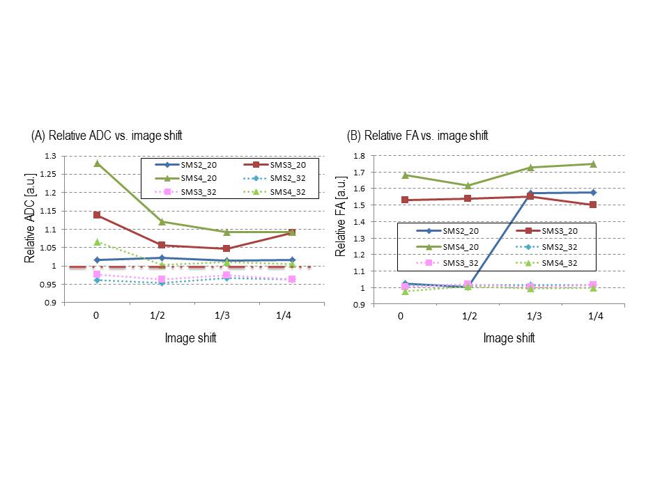
Effect of SMS factor on DTI metrics: SMS factors 3 and 4 of the 20ch head coil overestimated ADC (Fig. 4A) and FA (Fig. 4B). Almost all SMS factors on the 32ch and SMS factor 2 on the 20ch head coil showed values close to conventional DTI, except when image shifts of SMS factor 2 on 20ch were 1/3 or 1/4.
Clinical feasibility of SMS-EPI DTI: Figure 5 shows the results of visual inspection by the two experienced neuroradiologists. With SMS factors 2 and 3, imaging shifts (>0) with the 32ch head coil were judged clinically feasible and can be substituted on conventional DTI. For the 20ch head coil, only SMS factor 2 was judged clinically feasible, except when imaging shift was 1/3.
LIMITATIONS
Only one healthy volunteer was investigated in this study. For better statistical reliability, further investigation with a larger number of participants is needed. Further investigations are needed before clinical applications can be countenanced.CONCLUSION
SMS factors 2 and 3 with imaging shifts (>1/2) on 32ch were considered clinically feasible and can be substituted on conventional DTI. SMS factor 2 with imaging shifts 0, 1/2, 1/4 on the 20ch head coil were judged as clinically feasible.Acknowledgements
The prototype simultaneous multi-slice echo planar imaging acquisition technique for diffusion tensor imaging was provided from Siemens GmbH.References
[1] Setsompop et al., MRM, 2012; 67: 1210-1224, [2] Diffusion toolkit, https://www.nitrc.org/projects/trackvis/, [3] Oishi et al., Nuroimage 2009; 46(2): 486-499, [4] Diffeomap, http://www.mri-resource.kennedykrieger.org/software , [5] S. B. Reeder et al., Magnetic Resonance in Medicine, vol. 54, 2005, pp. 748-754.Figures